Pulsed-Wave Doppler Echocardiography
By measuring Doppler shift, modern ultrasonographs quantify blood
flow velocities. Doppler shift is the shift in frequency of a wave when the source
of the wave is moving (in this case, the wave reflected by moving red blood cells).
When pulses of sound are used (pulsed-wave [PW] Doppler), the operator defines a
small area (termed "sample volume") anywhere in the two-dimensional sector scan,
and the ultrasonograph automatically converts the Doppler data in that sample volume
to a display of the real-time blood flow velocities ( Fig.
33-3
). Thus, PW Doppler defines blood flow velocities and their location
within the heart and great vessels.
However, two important limitations apply. First, Doppler shift
is proportional to the cosine of the angle
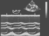 Figure 33-1
M-mode transesophageal echocardiogram of a normal aortic
valve. For reference, a single frame (stop action) of the two-dimensional cross
section is shown at the top right of the figure. The dotted
vertical line through the two-dimensional echocardiogram depicts the single
line of sampling provided by the M-mode echocardiogram over time (the horizontal
axis for the lower two thirds of the figure). The electrocardiogram defines systole
and diastole. Note in the middle of the M-mode image the three tilted
rectangles connected by the slightly undulating line.
These rectangles and lines are formed by the motion of the leaflets of the aortic
valve as they open and close during the cardiac cycles shown. From top to bottom
in this M-mode echocardiogram, the structures indicated by the white
lines are the posterior wall of the left atrium (just under the electrocardiogram),
the posterior wall of the aortic annulus, the aortic valve (as described above),
the anterior wall of the aortic annulus, a pulmonary artery catheter, and the myocardium
of the right ventricular outflow tract. (From Cahalan MK: Intraoperative
Transesophageal Echocardiography. An Interactive Text and Atlas. New York, Churchill
Livingstone, 1997.)
Figure 33-1
M-mode transesophageal echocardiogram of a normal aortic
valve. For reference, a single frame (stop action) of the two-dimensional cross
section is shown at the top right of the figure. The dotted
vertical line through the two-dimensional echocardiogram depicts the single
line of sampling provided by the M-mode echocardiogram over time (the horizontal
axis for the lower two thirds of the figure). The electrocardiogram defines systole
and diastole. Note in the middle of the M-mode image the three tilted
rectangles connected by the slightly undulating line.
These rectangles and lines are formed by the motion of the leaflets of the aortic
valve as they open and close during the cardiac cycles shown. From top to bottom
in this M-mode echocardiogram, the structures indicated by the white
lines are the posterior wall of the left atrium (just under the electrocardiogram),
the posterior wall of the aortic annulus, the aortic valve (as described above),
the anterior wall of the aortic annulus, a pulmonary artery catheter, and the myocardium
of the right ventricular outflow tract. (From Cahalan MK: Intraoperative
Transesophageal Echocardiography. An Interactive Text and Atlas. New York, Churchill
Livingstone, 1997.)
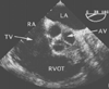 Figure 33-2
Short-axis two-dimensional cross section of a normal
aortic valve (AV). This midesophageal short-axis view of the AV reveals the morphology
of the three cusps of this normal valve. LA, left atrium; RA, right atrium; RVOT,
right ventricular outflow tract; TV, tricuspid valve. (From Cahalan MK:
Intraoperative Transesophageal Echocardiography. An Interactive Text and Atlas.
New York, Churchill Livingstone, 1997.)
Figure 33-2
Short-axis two-dimensional cross section of a normal
aortic valve (AV). This midesophageal short-axis view of the AV reveals the morphology
of the three cusps of this normal valve. LA, left atrium; RA, right atrium; RVOT,
right ventricular outflow tract; TV, tricuspid valve. (From Cahalan MK:
Intraoperative Transesophageal Echocardiography. An Interactive Text and Atlas.
New York, Churchill Livingstone, 1997.)
between the ultrasound beam and the direction of the blood cells. If the cells are
moving directly parallel to the ultrasound beam, the angle is zero, and the cosine
of zero is one. Thus, at an angle of zero, Doppler echocardiography provides a true
estimate of blood flow velocity. At
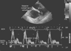 Figure 33-3
Pulsed-wave Doppler echocardiogram of the main pulmonary
artery (MPA). At the top of the echocardiogram is a still-frame image of the two-dimensional
cross section used to position the Doppler sample volume (the round
white sphere). On the bottom two thirds of the echocardiogram is the
display in white of the instantaneous blood flow velocities (vertical axis) versus
time (horizontal axis) occurring in that sample volume. The electrocardiogram provides
timing, and the bold horizontal line is the baseline
(zero flow) for the flow velocities. Flow velocities above this line are positive
(i.e., toward the transducer) to a maximum of 68 cm/sec. Flow below this line is
negative (i.e., away from the transducer) to a maximum of -14 cm/sec. (From
Cahalan MK: Intraoperative Transesophageal Echocardiography. An Interactive Text
and Atlas. New York, Churchill Livingstone, 1997.)
Figure 33-3
Pulsed-wave Doppler echocardiogram of the main pulmonary
artery (MPA). At the top of the echocardiogram is a still-frame image of the two-dimensional
cross section used to position the Doppler sample volume (the round
white sphere). On the bottom two thirds of the echocardiogram is the
display in white of the instantaneous blood flow velocities (vertical axis) versus
time (horizontal axis) occurring in that sample volume. The electrocardiogram provides
timing, and the bold horizontal line is the baseline
(zero flow) for the flow velocities. Flow velocities above this line are positive
(i.e., toward the transducer) to a maximum of 68 cm/sec. Flow below this line is
negative (i.e., away from the transducer) to a maximum of -14 cm/sec. (From
Cahalan MK: Intraoperative Transesophageal Echocardiography. An Interactive Text
and Atlas. New York, Churchill Livingstone, 1997.)
all other angles the cosine is less than one, which results in less Doppler shift
and an underestimation of flow velocity. Clinically, angles less than 15 degrees
(the cosine is almost one) are insignificant in the estimation of velocity, whereas
angles exceeding 20 degrees markedly attenuate the Doppler shift and thus necessitate
caution in interpreting such data. This relationship is best stated in the following
Doppler equation:
V = C · (Fd
)/(2F0
·
cos theta)
where V is the velocity to be measured, C is the speed of sound in tissue (a constant),
Fd
is the Doppler shift in ultrasound frequency measured by the ultrasound
machine, F0
is the frequency of the transducer, and cos theta is the
cosine of the angle between the direction of the blood flow and the ultrasound waves.
Second, the maximum velocity of blood flow that can be unambiguously measured is
defined by the Nyquist limit. The Nyquist limit is directly related to the ultrasound
frequency and the pulse repetition frequency. The pulse repetition frequency is
the number of pulses of ultrasound emitted per second. Because an emitted pulse
must return before the next pulse is emitted in PW Doppler, an increase in depth
of the ultrasound scan decreases the pulse repetition frequency and the Nyquist limit.
Thus, the depth of the ultrasound scan is the principal determinant of the Nyquist
limit under the control of the operator. If the velocity of blood flow exceeds the
Nyquist limit, sudden apparent flow reversal ("aliasing") will be depicted ( Fig.
33-4
). Aliasing is analogous to the sudden apparent reversal of direction
in stagecoach wheels visible in old Westerns when the velocity of the wheel spokes
exceeds the frame
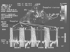 Figure 33-4
Pulsed-wave Doppler with aliasing at high velocities.
Pulsed-wave Doppler measurement of blood flow velocities in a mitral valve (MV)
orifice during four cardiac cycles is shown. At the top of the figure is a still-frame
image of the two-dimensional cross section used to position the Doppler sample volume
(the round white sphere). On the bottom two thirds
of the figure is the display in white of the instantaneous blood flow velocities
(vertical axis) versus time (horizontal axis) occurring in that sample volume. The
electrocardiogram provides timing, and the bold horizontal line
is the baseline (zero flow) for the flow velocities. Flow velocities above this
line are positive (i.e., toward the transducer) to a maximum of 183 cm/sec. Flow
velocities below this line are negative (i.e., away from the transducer) to a maximum
of -77 cm/sec. This tracing documents significant mitral regurgitation (the positive
systolic velocities) but does not measure the peak velocity of regurgitant flow because
it is beyond the Nyquist limit—the systolic velocities off the top of the scale
are said to alias, that is, they go off scale and wrap around into the domain of
negative velocities. LA, left atrium; LV, left ventricle. (From Cahalan
MK: Intraoperative Transesophageal Echocardiography. An Interactive Text and Atlas.
New York, Churchill Livingstone, 1997.)
Figure 33-4
Pulsed-wave Doppler with aliasing at high velocities.
Pulsed-wave Doppler measurement of blood flow velocities in a mitral valve (MV)
orifice during four cardiac cycles is shown. At the top of the figure is a still-frame
image of the two-dimensional cross section used to position the Doppler sample volume
(the round white sphere). On the bottom two thirds
of the figure is the display in white of the instantaneous blood flow velocities
(vertical axis) versus time (horizontal axis) occurring in that sample volume. The
electrocardiogram provides timing, and the bold horizontal line
is the baseline (zero flow) for the flow velocities. Flow velocities above this
line are positive (i.e., toward the transducer) to a maximum of 183 cm/sec. Flow
velocities below this line are negative (i.e., away from the transducer) to a maximum
of -77 cm/sec. This tracing documents significant mitral regurgitation (the positive
systolic velocities) but does not measure the peak velocity of regurgitant flow because
it is beyond the Nyquist limit—the systolic velocities off the top of the scale
are said to alias, that is, they go off scale and wrap around into the domain of
negative velocities. LA, left atrium; LV, left ventricle. (From Cahalan
MK: Intraoperative Transesophageal Echocardiography. An Interactive Text and Atlas.
New York, Churchill Livingstone, 1997.)
rate of the movie camera. Typically, aliasing of PW Doppler occurs at blood flow
velocities of 0.8 to 1.0 m/sec. Normal flow within the heart may reach 1.4 m/sec,
and pathologic flow, up to 6 m/sec. To measure these velocities, continuous-wave
(CW) Doppler is needed.
 |