|
|
|
|
|
|
|
|
|
|
|
|
|
|
|
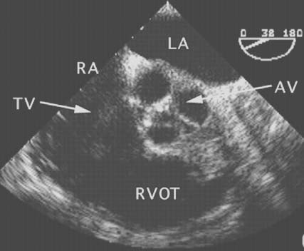
Figure 33-2
Short-axis two-dimensional cross section of a normal
aortic valve (AV). This midesophageal short-axis view of the AV reveals the morphology
of the three cusps of this normal valve. LA, left atrium; RA, right atrium; RVOT,
right ventricular outflow tract; TV, tricuspid valve. (From Cahalan MK:
Intraoperative Transesophageal Echocardiography. An Interactive Text and Atlas.
New York, Churchill Livingstone, 1997.)
|
|
|
|
|
|
|
|
|
|
|
|
|
|