|
|
|
|
|
|
|
|
|
|
|
|
|
|
|
As the flow-directed, balloon-tipped PAC is floated from a central vein to its proper position in the pulmonary artery, characteristic pressure waveforms are recorded (see Fig. 32-29 ). When the catheter tip reaches the superior vena cava or right atrium, a CVP waveform should be observed with its a, c, and v waves and low mean pressure value (see Table 32-7 ). At this point, the PAC balloon is inflated, and the catheter is advanced until it crosses the tricuspid valve to record right ventricular pressure. This waveform is recognized by the sudden increase in systolic pressure, the wide pulse pressure, and the low diastolic pressure that approximates CVP. The PAC next enters the right ventricular outflow tract and floats across the pulmonic valve into the main pulmonary artery. Often, this passage is heralded by arrhythmias, especially premature ventricular beats, as the balloon-tipped catheter strikes the right ventricular infundibulum. PAP is characterized by the step-up in diastolic pressure recorded as the catheter crosses the pulmonic valve. In the absence of pulmonic valve stenosis, pulmonary artery systolic pressure closely approximates right ventricular systolic pressure, but pulmonary artery diastolic pressure generally exceeds right ventricular diastolic pressure.
On occasion, it may be difficult to distinguish right ventricular pressure from PAP, particularly if only the numeric values for these pressures are examined. However, careful observation of the pressure waveforms, with a focus on the diastolic pressure contours, will allow
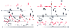 Figure 32-32
Temporal relationships between normal systemic arterial
pressure (ART), pulmonary artery pressure (PAP), central venous pressure (CVP), and
pulmonary artery wedge pressure (PAWP). Note that the PAWP a-c and v waves appear
to occur later in the cardiac cycle than do their counterparts on the right side
of the heart seen in the CVP trace. The ART scale is on the left,
and the PAP, CVP, and PAWP scales are on the right. (Redrawn from Mark JB:
Atlas of Cardiovascular Monitoring. New York, Churchill Livinstone, 1998, Fig.
3-3.)
Figure 32-32
Temporal relationships between normal systemic arterial
pressure (ART), pulmonary artery pressure (PAP), central venous pressure (CVP), and
pulmonary artery wedge pressure (PAWP). Note that the PAWP a-c and v waves appear
to occur later in the cardiac cycle than do their counterparts on the right side
of the heart seen in the CVP trace. The ART scale is on the left,
and the PAP, CVP, and PAWP scales are on the right. (Redrawn from Mark JB:
Atlas of Cardiovascular Monitoring. New York, Churchill Livinstone, 1998, Fig.
3-3.)
Under normal conditions, the PAP upstroke slightly precedes the radial artery pressure upstroke ( Fig. 32-32 ) because of the longer duration of left ventricular isovolumic contraction, as well as the transmission time of the central aortic pressure upstroke to the downstream radial artery recording site. Although the PAP upstroke precedes the radial artery pressure upstroke by 50 milliseconds, peak PAP precedes peak radial artery pressure by only 10 milliseconds.[429] As a practical matter, the pulmonary and systemic arterial pressure waveforms appear to overlap on the bedside monitor, with these pressures rising, peaking, and falling at approximately the same points in time (see Fig. 32-32 ). Understanding these temporal relationships is critically important if one is to properly interpret abnormal pulmonary artery and wedge pressure waveforms, particularly when tall v waves are present in these traces (see later).
The PAC, with balloon inflated, finally reaches the wedge position. As noted earlier, wedge pressure is an indirect measurement of pulmonary venous pressure and left atrial pressure and should therefore resemble these pressure waveforms. Consequently, the PAWP waveform may be identified as a venous pressure trace that displays characteristic a and v waves and x and y descents. However, because of the pulmonary vascular bed interposed between the PAC tip and the left atrium, wedge pressure is a delayed representation of left atrial pressure.[423] [430] On average, 160 milliseconds is required for the left atrial pressure pulse to traverse the pulmonary veins, capillaries, arterioles, and arteries. As a consequence of this delay, the wedge pressure a wave appears to follow the ECG R wave in early ventricular systole (see Fig. 32-32 ). However, the a wave is an end-diastolic pressure event produced by left atrial contraction. Recognition of
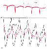 Figure 32-33
Pulmonary artery wedge pressure (PAWP) is a damped, delayed
reflection of left atrial pressure (LAP). The tall regurgitant v waves (v) caused
by severe mitral regurgitation are seen clearly in the LAP trace but appear delayed
and smaller in magnitude in the PAWP trace. (Redrawn from Mark JB: Atlas
of Cardiovascular Monitoring. New York, Churchill Livingstone, 1998, Fig. 4-4.)
Figure 32-33
Pulmonary artery wedge pressure (PAWP) is a damped, delayed
reflection of left atrial pressure (LAP). The tall regurgitant v waves (v) caused
by severe mitral regurgitation are seen clearly in the LAP trace but appear delayed
and smaller in magnitude in the PAWP trace. (Redrawn from Mark JB: Atlas
of Cardiovascular Monitoring. New York, Churchill Livingstone, 1998, Fig. 4-4.)
Not only is wedge pressure a delayed representation of left atrial pressure, but it is also a damped reflection of phasic atrial pressure waves. The amount of damping is variable, but when left atrial pressure waves are prominent, the pressure peaks may be significantly underestimated by the wedge trace ( Fig. 32-33 ). Even though the wedge pressure waveform will always appear to be a damped, delayed version of left atrial pressure, the mean pressure recorded from these two sites should be similar under most circumstances.
To recognize prominent a or v waves in the wedge pressure trace, it is not always necessary to inflate the PAC balloon and obtain a wedge position ( Fig. 32-34 ). Because the wedge pressure records pressure waves transmitted in retrograde fashion from the left atrium, these waves will normally sum with antegrade PAP waves produced by right ventricular ejection. The PAP trace thus becomes a composite wave reflecting both retrograde and antegrade components. Tall left atrial a or v waves will distort the normal PAP waveform appearance, with the a wave inscribed at the onset of systolic upstroke and the v wave distorting the dicrotic notch (see Fig. 32-34 ).[423] [431] Once these waves are identified by wedging the PAC and comparing the pulmonary artery and wedge pressure traces, it is convenient and perhaps more prudent to "follow" the wedge pressure a and v waves in the unwedged
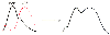 Figure 32-34
Tall left atrial pressure (LAP) a and v waves transmitted
in a retrograde direction through the pulmonary vasculature distort the antegrade
pulmonary artery pressure (PAP) waveform. The LAP a wave distorts systolic upstroke,
and the v wave distorts the dicrotic notch. (Redrawn from Mark JB: Atlas
of Cardiovascular Monitoring. New York, Churchill Livingstone, 1998, Fig. 4-10.)
Figure 32-34
Tall left atrial pressure (LAP) a and v waves transmitted
in a retrograde direction through the pulmonary vasculature distort the antegrade
pulmonary artery pressure (PAP) waveform. The LAP a wave distorts systolic upstroke,
and the v wave distorts the dicrotic notch. (Redrawn from Mark JB: Atlas
of Cardiovascular Monitoring. New York, Churchill Livingstone, 1998, Fig. 4-10.)
In summary, PAWP measured with a balloon-tipped PAC provides a delayed, damped estimate of left atrial pressure by measuring the pressure where flow resumes at a pulmonary venous junction point near the left atrium. Mean wedge pressure will always be lower than mean PAP; otherwise, blood would not flow in an antegrade direction. The wedge pressure waveform should display small a and v waves that should be identifiable if the pressure trace is displayed with sufficient gain and resolution on the monitor screen.
|
|
|
|
|
|
|
|
|
|
|
|
|