|
|
|
|
|
|
|
|
|
|
|
|
|
|
|
In 1970, Swan, Ganz, and colleagues introduced pulmonary artery catheterization into clinical practice for hemodynamic assessment of patients with acute myocardial infarction.[313] [314] [315] [316] [317] These catheters allowed accurate measurement of important cardiovascular physiologic variables at the bedside, and their popularity soared over the next 2 decades as they were used in an increasing number of critically ill and surgical patients. By the middle 1990s, estimated annual pulmonary artery catheter (PAC) sales in the United States approached 2 million catheters, with an estimated cost associated with their use in excess of $2 billion each year.[318]
The PAC provides measurements of several hemodynamic variables that many clinicians, including experts in intensive care, cannot accurately predict from standard clinical signs and symptoms.[319] [320] However, despite this evidence that PACs provide cardiovascular measurements that supplement or correct clinical observations, it remains uncertain whether PAC monitoring leads to improved patient outcome.[321] [322] [323] [324] [325] Before considering this issue, we will review techniques for catheterization, complications associated with PAC use, the physiologic basis for PAC monitoring, interpretation of normal and pathologic PAC waveforms, and the relationship between PAC-derived measures of left ventricular filling pressure and left ventricular preload.
PACs can be placed from any of the central venous cannulation sites described earlier, but the right internal jugular vein is preferred because it provides the most direct route to the right heart chambers and thereby usually leads to successful catheterization. The procedure is conducted as already described for central venous cannulation, until the step where the venous catheter is to be inserted. At this point, a slightly larger skin nick is made to accommodate the large-bore (7.5 to 9.0 French) introducer sheath. This introducer has a hemostasis valve at its outer end, through which the PAC will be inserted, and a sidearm extension that allows continuous central venous access for fluid and drug administration. A tapered-tip, stiff dilator stylet is placed into the introducer sheath to facilitate passage of this large cannula over the guidewire from the skin, through the subcutaneous tissues, into the vein. Utmost care must be exercised when introducing this large dilator-cannula assembly by advancing it only to the depth required to enter the vein and then threading the introducer cannula completely into the vein over the guidewire and dilator. Finally, the guidewire and dilator are removed, the sidearm extension is secured to an intravenous infusion set with a Luer-Lock connector, and the introducer sheath is sutured in place.
The standard PAC has a 7.0-, 7.5-, or 8.0-French circumference and is 110 cm in length, with these distances marked at 10-cm intervals. It contains four separate internal lumens. One leads to the distal port at the catheter tip and is used for PAP monitoring. The second leads to a proximal port, located approximately 30 cm from the catheter tip, and is intended for CVP monitoring and fluid and drug administration. The third lumen leads to a balloon near the catheter tip, and the fourth contains fine wires leading to a temperature thermistor, just proximal to the balloon. This thermistor is used to monitor pulmonary artery blood temperature as part of the thermodilution method for cardiac output monitoring (see later).
The final steps in placing a PAC require the aid of a skilled assistant to help prepare the catheter and attach it properly to pressure monitoring transducers. The physician performing the catheterization removes the PAC from its package and inserts it through a sterile plastic sheath while being careful to not damage the balloon in this process.[326] This sheath or sleeve covers a length of the PAC residing outside the patient and thereby allows minor manipulations in PAC position during the monitoring period while attempting to maintain catheter sterility. The assistant then attaches the distal and proximal port hubs to the pressure monitoring system that will allow these waveforms to be displayed on the bedside monitor. These ports are flushed to ensure proper function and expel the air from these catheter lumens. The balloon is inflated with a 1.5-mL volume-limited syringe packaged with the PAC, and the balloon is tested by filling it completely with 1.5 mL of air to make certain that there are no leaks and that the balloon inflates symmetrically without obstructing the opening of the distal end of the lumen.[327]
The balloon should always be inflated with air, not liquid. The air-filled balloon at the tip of the catheter serves to carry the catheter forward with blood flow through the right heart chambers and helps "float" the catheter to its proper position in the pulmonary artery, much like the spinnaker on a sailboat is driven by the trailing wind.
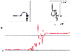 Figure 32-28
Bedside testing of the pulmonary artery catheter-pressure
transducer system. With the tip of the catheter held at the level of the transducer
and the heart, a pressure of 0 mm Hg is recorded. The catheter tip is raised to
create a 30-cm vertical fluid column, and the resulting pressure display should equal
22 mm Hg (equivalent to 30 cm H2
O). (Redrawn from Mark JB: Atlas
of Cardiovascular Monitoring. New York, Churchill Livingstone, 1998, Fig. 3-4.)
Figure 32-28
Bedside testing of the pulmonary artery catheter-pressure
transducer system. With the tip of the catheter held at the level of the transducer
and the heart, a pressure of 0 mm Hg is recorded. The catheter tip is raised to
create a 30-cm vertical fluid column, and the resulting pressure display should equal
22 mm Hg (equivalent to 30 cm H2
O). (Redrawn from Mark JB: Atlas
of Cardiovascular Monitoring. New York, Churchill Livingstone, 1998, Fig. 3-4.)
A simple maneuver should now be performed to check for proper assembly and function of the PAC monitoring system.[330] [331] When the tip of the PAC is held near heart level, the recorded pressure should be 0 mm Hg, thereby confirming that the transducer was correctly adjusted to the level of the heart at the beginning of the procedure. The catheter tip is then raised up straight to create
 Figure 32-29
Characteristic waveforms recorded during passage of a
pulmonary artery catheter. Right atrial pressure resembles a central venous pressure
waveform and displays a, c, and v waves. Right ventricular pressure shows a higher
systolic pressure than seen in the right atrium, although the end-diastolic pressures
are equal in these two chambers. Pulmonary artery pressure shows a diastolic step-up
when compared with ventricular pressure. Note also that right ventricular pressure
increases during diastole whereas pulmonary artery pressure decreases during diastole
(shaded boxes). Pulmonary artery wedge pressure
has a morphology similar to that of right atrial pressure, although the a-c and v
waves appear later in the cardiac cycle relative to the electrocardiogram. (Redrawn
from Mark JB: Atlas of Cardiovascular Monitoring. New York, Churchill Livingstone,
1998, Fig. 3-1.)
Figure 32-29
Characteristic waveforms recorded during passage of a
pulmonary artery catheter. Right atrial pressure resembles a central venous pressure
waveform and displays a, c, and v waves. Right ventricular pressure shows a higher
systolic pressure than seen in the right atrium, although the end-diastolic pressures
are equal in these two chambers. Pulmonary artery pressure shows a diastolic step-up
when compared with ventricular pressure. Note also that right ventricular pressure
increases during diastole whereas pulmonary artery pressure decreases during diastole
(shaded boxes). Pulmonary artery wedge pressure
has a morphology similar to that of right atrial pressure, although the a-c and v
waves appear later in the cardiac cycle relative to the electrocardiogram. (Redrawn
from Mark JB: Atlas of Cardiovascular Monitoring. New York, Churchill Livingstone,
1998, Fig. 3-1.)
The PAC is inserted through the hemostasis valve of the introducer to a depth of 20 cm, or approximately 5 cm beyond the tip of the introducer sheath. A characteristic CVP waveform must be identified to confirm that the PAC tip is in the vena cava or right atrium. The gentle curvature of the PAC should be oriented to point just leftward of the sagittal plane (the 11-o'clock position as viewed from the patient's head). This orientation will facilitate catheter passage through the anteromedially located tricuspid valve. The balloon is inflated, and the catheter is advanced into the right atrium, through the tricuspid valve into the right ventricle, through the pulmonic valve into the pulmonary artery, and finally into the wedge position. Characteristic waveforms from each of these locations confirm proper catheter passage and placement ( Fig. 32-29 ).
After PAWP is measured, the balloon is deflated, and the PAP waveform should reappear for continuous monitoring. Wedge pressure may be obtained periodically as needed by reinflating the balloon with 1.5 mL of air to allow the catheter to float distally until pulmonary artery occlusion again occurs. The clinician must recognize that the PAC tip advances to a smaller pulmonary artery each time that the balloon is inflated to measure wedge pressure. Similarly, the PAC should return to its monitoring position in the proximal portion of the pulmonary artery after the balloon is deflated. In some patients, the PAC may migrate distally even without balloon inflation. This problem is more common during cardiopulmonary bypass because of the repeated cardiac manipulations and temperature changes that alter the stiffness of the catheter.[332] Consequently, proper catheter position must be ensured throughout the monitoring period by frequent observation of the PAP waveform. If a PAWP trace appears without balloon inflation or with only partial
From a right internal jugular vein puncture site, the right atrium should be reached when the PAC is inserted 20 to 25 cm, the right ventricle at 30 to 35 cm, the pulmonary artery at 40 to 45 cm, and the wedge position at 45 to 55 cm. When other sites are chosen for catheter placement, additional distance is required, typically an additional 5 to 10 cm from the left internal jugular and left and right external jugular veins, an additional 15 cm from the femoral veins, and an additional 30 to 35 cm from the antecubital veins.[331] These distances serve only as a rough guide, and waveform morphology must always be verified from the monitor display and catheter position confirmed with a chest radiograph. The tip of the PAC should be within 2 cm of the cardiac silhouette on a standard anteroposterior chest film.[334]
Keeping in mind these typical distances helps avoid complications caused by unintended catheter loops and knots within the heart. If a right ventricular waveform is not observed after inserting the catheter 40 cm, coiling in the right atrium is likely. Similarly, if a pulmonary artery waveform is not observed after inserting the catheter 50 cm, coiling in the right ventricle has probably occurred. The balloon should be deflated, the catheter withdrawn to 20 cm, and the PAC floating sequence repeated.
Although the right internal jugular vein is the primary site for pulmonary artery catheterization, failure to achieve venous cannulation or other medical and surgical considerations may require the use of alternative sites. For cardiac surgery, the left internal jugular vein is often the second choice because use of the subclavian veins poses a problem unique to this setting. After sternotomy and sternal retraction, a PAC inserted by the subclavian route may be compressed and kinked under the clavicle, thus making it impossible to withdraw or advance the catheter should this be required during the course of surgery.[335]
Regardless of the intended surgical procedure, whenever one plans to insert a PAC through the left internal jugular, subclavian, and particularly the external jugular veins, the more tortuous course of these vessels may make it difficult to advance the large-bore introducer sheath fully into the vessel. This is not a problem as long as the introducer cannula is withdrawn slightly to maintain a position in the vein that allows unimpeded intravenous infusion and free aspiration of venous blood. Often, the introducer cannula needs to remain in this partially inserted position while the PAC is introduced through its hemostasis valve. Because the PAC has a more flexible, smaller tip than the introducer does, the PAC might advance easily and completely into the central vein and right atrium. The PAC balloon may then be inflated and the procedure completed in normal fashion. With the PAC in proper position, the introducer sheath can usually be fully guided into the central vein and sutured in place because the PAC has served, in essence, as a more effective "guidewire." Occasionally, the introducer sheath must remain partially or completely withdrawn to avoid acute angulation and kinking of the PAC as it leaves the sheath and enters the central vein.[335] [336] [337]
After successful venous cannulation, when attempts to advance the PAC to the right ventricle prove difficult, the physician should consider whether this difficulty is due to abnormal venous anatomy. The most common abnormality of the systemic veins is persistence of the left superior vena cava, which is present in approximately 0.1% to 0.2% of the general population and 2% to 9% of patients with other forms of congenital heart disease.[338] [339] [340] [341] [342] [343] A persistent left superior vena cava descends along the left mediastinum and empties into the coronary sinus, which is dilated as a consequence of the abnormal venous drainage pattern. Occasionally, the condition is recognized radiographically by an abnormal appearance of the left side of the mediastinum and cardiac border or an abnormal pulsation of the left internal jugular vein. However, because of the lack of physiologic consequences of this abnormal venous anatomy, virtually all cases are asymptomatic and discovered incidentally at the time of attempted central venous or pulmonary artery catheterization. In patients with this anomaly, PAC placement may be difficult or impossible as a result of the tortuous course that the catheter must follow from the vena cava through the coronary sinus to reach the right heart chambers. Because a normal right superior vena cava is present in most of these patients, the anomaly is recognized only when attempted PAC placement proceeds from a left-sided vein. More rarely, difficult PAC placement is encountered during attempted right-sided venous cannulation because the right superior vena cava is absent as well. In these cases, the right internal jugular vein joins the persistent left superior vena cava by a bridging innominate vein. A rare form of atrial septal defect termed unroofed coronary sinus may also be encountered in patients with these venous abnormalities. This congenital defect provides a site for coronary sinus-left atrial communication, with the potential for a PAC to enter the left atrium and systemic circulation through this defect.[343] With any of these venous anomalies, PAC advancement into the coronary sinus may disclose an unexpected pressure waveform, coronary sinus pressure. This waveform has characteristics of both the downstream right atrial pressure and an indirectly transmitted, dampened left ventricular pressure waveform. Recognition requires that the physician have a clear appreciation for the expected waveforms encountered by the PAC as right heart catheterization proceeds.
A few additional points might aid successful positioning of the PAC. The air-filled balloon tends to float to nondependent regions as it passes through the heart into the pulmonary vasculature. Consequently, positioning a patient head down will aid flotation past the tricuspid valve, and tilting the patient onto the right side and head-up positioning will aid flotation out of the right ventricle and may reduce the frequency of malignant ventricular arrhythmias during catheterization. [344]
|
|
|
|
|
|
|
|
|
|
|
|
|