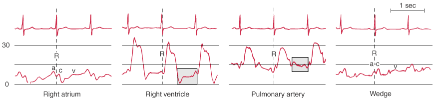
Figure 32-29
Characteristic waveforms recorded during passage of a
pulmonary artery catheter. Right atrial pressure resembles a central venous pressure
waveform and displays a, c, and v waves. Right ventricular pressure shows a higher
systolic pressure than seen in the right atrium, although the end-diastolic pressures
are equal in these two chambers. Pulmonary artery pressure shows a diastolic step-up
when compared with ventricular pressure. Note also that right ventricular pressure
increases during diastole whereas pulmonary artery pressure decreases during diastole
(shaded boxes). Pulmonary artery wedge pressure
has a morphology similar to that of right atrial pressure, although the a-c and v
waves appear later in the cardiac cycle relative to the electrocardiogram. (Redrawn
from Mark JB: Atlas of Cardiovascular Monitoring. New York, Churchill Livingstone,
1998, Fig. 3-1.)