Physiologic Considerations for Pulmonary Artery Catheter
Monitoring: Prediction of Left Ventricular Filling Pressure
Pulmonary artery catheterization is performed in critically ill
patients to measure a range of hemodynamic variables, including cardiac output, mixed
venous oxygen saturation, and most importantly, pulmonary artery diastolic and wedge
pressures. These pressure measurements are used to estimate left ventricular filling
pressure and help guide fluid and vasoactive drug administration when clinical signs,
symptoms, or other monitored variables are thought to be inadequate or unreliable.
When a PAC floats to the wedge position, the inflated balloon
at its tip isolates the distal pressure monitoring orifice from upstream PAP. Blood
flow ceases between the catheter tip and a junction point where pulmonary veins draining
the occluded pulmonary vascular region join other veins in which blood still flows
toward the left atrium ( Fig. 32-30
).
A continuous static column of blood now connects the wedged PAC tip to this junction
point
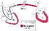 Figure 32-30
Pulmonary artery wedge pressure measurement creates a
static column of blood connecting the catheter tip to a junction point where flow
resumes in the pulmonary veins (PV) near the left atrium (LA). LV, left ventricle;
PA, pulmonary artery; RA, right atrium; RV, right ventricle. (Redrawn from
Mark JB: Atlas of Cardiovascular Monitoring. New York, Churchill Livingstone, 1998,
Fig. 4-1.)
Figure 32-30
Pulmonary artery wedge pressure measurement creates a
static column of blood connecting the catheter tip to a junction point where flow
resumes in the pulmonary veins (PV) near the left atrium (LA). LV, left ventricle;
PA, pulmonary artery; RA, right atrium; RV, right ventricle. (Redrawn from
Mark JB: Atlas of Cardiovascular Monitoring. New York, Churchill Livingstone, 1998,
Fig. 4-1.)
in the pulmonary veins near the left atrium. Thus, wedging the PAC functionally
extends the catheter tip to measure the pressure at the point at which blood flow
resumes on the venous side of the pulmonary circuit.[422]
Because resistance to flow in the large pulmonary veins is negligible, PAWP provides
an accurate, indirect measurement of both pulmonary venous pressure and left atrial
pressure.[120]
[423]
Pulmonary artery diastolic pressure is often used as an alternative
to wedge pressure as an estimate of left ventricular filling pressure. Under normal
circumstances, resistance to pulmonary blood flow is low, and the pressure in the
pulmonary artery at the end of diastole equilibrates with downstream pressure in
the pulmonary veins and left atrium.[424]
[425]
[426]
[427]
From
a monitoring standpoint, pulmonary artery diastolic pressure has the added
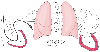 Figure 32-31
The pulmonary artery catheter tip must be wedged in lung
zone 3 to provide an accurate measure of pulmonary venous (Pv
) or left
atrial (LA) pressure. When alveolar pressure (PA
) rises above Pv
in lung zone 2 or above pulmonary arterial pressure (Pa
) in lung zone
1, wedge pressure will reflect alveolar pressure rather than intravascular pressure.
LV, left ventricle; PA, pulmonary artery; RA, right atrium; RV, right ventricle.
(Redrawn from Mark JB: Atlas of Cardiovascular Monitoring. New York, Churchill
Livingstone, 1998, Fig. 6-10.)
Figure 32-31
The pulmonary artery catheter tip must be wedged in lung
zone 3 to provide an accurate measure of pulmonary venous (Pv
) or left
atrial (LA) pressure. When alveolar pressure (PA
) rises above Pv
in lung zone 2 or above pulmonary arterial pressure (Pa
) in lung zone
1, wedge pressure will reflect alveolar pressure rather than intravascular pressure.
LV, left ventricle; PA, pulmonary artery; RA, right atrium; RV, right ventricle.
(Redrawn from Mark JB: Atlas of Cardiovascular Monitoring. New York, Churchill
Livingstone, 1998, Fig. 6-10.)
advantage of being available for continuous monitoring, in contrast to wedge pressure,
which can be measured only intermittently when the PAC balloon is inflated.
For pulmonary artery diastolic or wedge pressure to be a valid
estimate of left ventricular filling pressure, a continuous, static column of blood
must connect the tip of the wedged catheter and the draining pulmonary venous radicle.
At the microcirculatory level, this connecting channel consists of thin pulmonary
capillaries, which are subject to extramural compressive forces exerted by the surrounding
alveoli. West and colleagues described a physiologic model of the pulmonary vasculature
consisting of three zones that are based on the gravitationally determined relationships
between PAP, pulmonary venous pressure, and alveolar pressure.[428]
This lung zone model provides useful insight into conditions when the PAC may not
provide accurate estimates of left ventricular filling pressure.
In lung physiologic zones 1 and 2, alveolar pressure can exceed
pulmonary venous pressure (zone 2) or both PAP and pulmonary venous pressure (zone
1) ( Fig. 32-31
). A PAC
positioned in these lung zones will be influenced by alveolar pressure, and the resultant
pressure will bear little relationship to the downstream pulmonary venous pressure
or left ventricular filling pressure. Under these circumstances, alveolar or airway
pressure is being monitored rather than the intended vascular pressure in the left
atrium or ventricle. Fortunately, in most clinical settings in which a PAC is used,
patients are supine, which favors the creation of zone 3 conditions, and radiographic
studies confirm that PAC tips lie below the level of the left atrium under these
clinical conditions.[346]
However, when patients
are placed in the lateral or semiupright position, considerable nondependent portions
of their lungs may exhibit zone 2 behavior. In general, zones 1 and 2 become more
extensive when left atrial pressure is low, when the PAC tip is located vertically
above the left atrium, or when alveolar pressure is high. Catheters that are wedged
outside zone 3 may be suspected when the normal phasic wedge pressure a and v waves
are absent, when wedge pressure varies markedly with the respiratory cycle, and when
mean wedge pressure exceeds pulmonary artery diastolic pressure because this should
never occur unless tall a or v waves are present in the wedge pressure trace.[120]