|
|
|
|
|
|
|
|
|
|
|
|
|
|
|
All modern volatile anesthetics, including desflurane and sevoflurane, depress contractile function in normal myocardium in vitro and in vivo.[1] Investigations conducted in the 1960s demonstrated that halothane causes dose-related depression of force-velocity relationships and Frank-Starling curves in isolated cardiac muscle preparations[2] [3] and intact, closed-chest dogs,[4] respectively. These findings supported clinical observations of circulatory depression during halothane anesthesia in humans.[5] [6] [7] [8] Enflurane [9] and isoflurane[10] produce direct negative inotropic effects, as indicated by decreases in maximal velocity of shortening, peak developed force, and maximal rate of force development during isotonic contraction in isolated feline papillary muscles. These reductions in intrinsic myocardial contractility by enflurane and isoflurane contribute to the cardiovascular depression observed with these agents in humans.[11] [12]
The relative degree of myocardial depression produced by different volatile anesthetics in vivo has been more difficult to establish because simultaneous alterations in systemic and pulmonary hemodynamics and autonomic nervous system activity often complicate assessment of left ventricular (LV) systolic function. Early studies using isovolumic and ejection-phase measures of myocardial contractility demonstrated that enflurane and halothane caused similar negative inotropic effects in dogs[13] [14] [15] [16] and humans.[11] These findings were subsequently confirmed using the slope (Ees ) of the LV end-systolic pressure-midaxis diameter relationship as a relatively heart rate- and load-independent index of the inotropic state in chronically instrumented dogs.[17] Equi-anesthetic concentrations of enflurane and halothane depressed myocardial contractility to similar degrees in vivo.[17] In contrast, experimental animal studies have repeatedly demonstrated that isoflurane produces less myocardial depression than does halothane or enflurane. Halothane caused larger reductions in the rate of increase of LV positive pressure (dP/dt) than did isoflurane when equi-anesthetic concentrations were directly compared in the presence and absence of autonomic nervous system function,[16] [18] [19] suggesting that differences in myocardial depression caused by these anesthetics occurred independent of autonomic nervous system activity. [16] Differences in the negative inotropic effects of halothane and isoflurane have been quantified using the slope (Mw ) of the regional preload recruitable stroke work (PRSW) relationship derived from a differentially loaded series of LV pressure-segment length diagrams.[20] [21] In these studies conducted in chronically instrumented dogs, isoflurane maintained contractility an average of 20% higher than with equi-minimum alveolar concentrations (MACs) of halothane.[20] [21] Differences in the relative degree of myocardial depression produced by halothane and isoflurane in humans have also been inferred using isovolumic and ejection-phase measures of contractile function.[12] [22] [23] The negative inotropic actions of isoflurane ( Fig. 7-1 ) and halothane are exacerbated by hypocalcemia,[24] calcium ion (Ca2+ ) channel blockers,[25] [26] [27] and β1 -adrenoceptor antagonists[25] and can be reversed by administration of exogenous Ca2+ ,[24] [28] [29] [30] cardiac phosphodiesterase fraction III inhibitors,[31] [32] β1 -adrenoceptor agonists,[1] Ca2+ channel agonists,[27] and myofilament Ca2+ sensitizers.[33] The differential effects of halothane and isoflurane on myocardial contractility in dogs are maintained during depression or augmentation of an inotropic state produced by these vasoactive drugs.[1]
Desflurane causes systemic and coronary hemodynamic effects that are remarkably similar to those produced by isoflurane.[34] Desflurane and isoflurane have been shown to depress myocardial function to equivalent degrees using isovolumic and ejection-phase measures of contractility in dogs,[16] [35] pigs,[36] and humans.[37] [38] These observations have been verified using end-systolic pressure-volume relationships and regional and global PRSW ( Fig. 7-2 ) in the presence and absence of autonomic nervous system activity in vivo.[39] [40] [41] However, the unique cardiovascular stimulation associated with rapid increases in inspired desflurane concentration in humans[42] may lead to transient increases in myocardial contractility resulting from augmentation of sympathetic nervous system tone.[43] [44] [45]
 Figure 7-1
End-systolic pressure-volume (top)
and stroke work versus end-diastolic volume relationships (bottom)
before (control 1 [C1
]); during 0.6, 0.9, and 1.2 minimum alveolar concentrations
(MACs); and after isoflurane (control 2 [C2
]) in an experiment in an open-chest
dog. (Adapted from Hettrick DA, Pagel PS, Warltier DC: Desflurane, sevoflurane,
and isoflurane impair canine left ventricular-arterial coupling and mechanical efficiency.
Anesthesiology 85:403–413, 1996.)
Figure 7-1
End-systolic pressure-volume (top)
and stroke work versus end-diastolic volume relationships (bottom)
before (control 1 [C1
]); during 0.6, 0.9, and 1.2 minimum alveolar concentrations
(MACs); and after isoflurane (control 2 [C2
]) in an experiment in an open-chest
dog. (Adapted from Hettrick DA, Pagel PS, Warltier DC: Desflurane, sevoflurane,
and isoflurane impair canine left ventricular-arterial coupling and mechanical efficiency.
Anesthesiology 85:403–413, 1996.)
The effects of sevoflurane on myocardial contractility are virtually indistinguishable from those produced by isoflurane in dogs.[46] [47] Another study conducted in pigs suggested that sevoflurane may produce less myocardial depression than equi-MACs of halothane.[48] Sevoflurane produces less myocardial depression than does enflurane in volunteers, as assessed by the heart rate-corrected velocity of circumferential fiber shortening versus the LV end-systolic wall stress relationship derived from echocardiography. [49] Sevoflurane decreased contractile function to approximately 40% to 45% of control values at 1.75 MAC in the presence and absence of autonomic nervous system tone using regional PRSW in dogs.[47] This magnitude of myocardial depression agrees with previous data for isoflurane and desflurane using an identical experimental model.[39] [40] Volatile anesthetics appear to depress the contractile state in normal ventricular myocardium
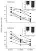 Figure 7-2
Preload recruitable stroke work (PRSW) slope for chronically
instrumented dogs during control and at 1.0 and 1.5 end-tidal minimum alveolar concentrations
(MAC) of desflurane (top) and isoflurane (bottom).
Insets depict percent changes from control values.
*, Significantly (P < .05) different from
controls; †, significantly (P < .05) different
from 1.0 MAC. (From Pagel PS, Kampine JP, Schmeling WT, Warltier DC: Influence
of volatile anesthetics on myocardial contractility in vivo: Desflurane versus isoflurane.
Anesthesiology 74:900–907, 1991.)
Figure 7-2
Preload recruitable stroke work (PRSW) slope for chronically
instrumented dogs during control and at 1.0 and 1.5 end-tidal minimum alveolar concentrations
(MAC) of desflurane (top) and isoflurane (bottom).
Insets depict percent changes from control values.
*, Significantly (P < .05) different from
controls; †, significantly (P < .05) different
from 1.0 MAC. (From Pagel PS, Kampine JP, Schmeling WT, Warltier DC: Influence
of volatile anesthetics on myocardial contractility in vivo: Desflurane versus isoflurane.
Anesthesiology 74:900–907, 1991.)
The effects of volatile anesthetics on LV systolic function in animal models or patients with LV dysfunction have not been comprehensively studied. Early in vitro studies demonstrated that isoflurane[10] and enflurane[50] caused larger reductions in maximal shortening velocity and the peak rate of force development in feline papillary muscles from failing hearts subjected to chronic pressure overload than in those from normal hearts. Halothane also produced more pronounced myocardial depression in ischemic than in normal myocardium.[51] [52] Isoflurane and halothane caused greater negative inotropic effects in ventricular myocardium obtained from cardiomyopathic hamsters than from normal hamsters ( Fig. 7-3 ).[53] These findings suggested that myocardial depression caused by volatile anesthetics in failing myocardium were accentuated, and they provided indirect evidence that patients with underlying contractile dysfunction might be more sensitive to the negative inotropic effects of
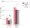 Figure 7-3
Comparison of the effects of halothane (left)
and isoflurane (right) on the isometric active force
(AF) of papillary muscles from healthy hamsters (red bars)
and those with cardiomyopathy (black and gray bars).
Probability values refer to between-group differences. *, Significantly (P
< .05) different from controls. (Adapted from Vivien B, Hanouz J-L, Gueugniaud
P-Y, et al: Myocardial effects of halothane and isoflurane in hamsters with hypertrophic
cardiomyopathy. Anesthesiology 87:1406–1416, 1997.)
Figure 7-3
Comparison of the effects of halothane (left)
and isoflurane (right) on the isometric active force
(AF) of papillary muscles from healthy hamsters (red bars)
and those with cardiomyopathy (black and gray bars).
Probability values refer to between-group differences. *, Significantly (P
< .05) different from controls. (Adapted from Vivien B, Hanouz J-L, Gueugniaud
P-Y, et al: Myocardial effects of halothane and isoflurane in hamsters with hypertrophic
cardiomyopathy. Anesthesiology 87:1406–1416, 1997.)
In experimental models of myocardial ischemia[51] or infarction,[54] volatile anesthetic-induced declines in contractile function were well tolerated and did not precipitate frank systolic dysfunction. Volatile anesthetics can exert important beneficial effects on mechanical function during myocardial ischemia and reperfusion injury. Volatile anesthetics reduce experimental myocardial infarct size,[55] [56] preserve metabolic and structural integrity during regional ischemia and reperfusion,[57] [58] [59] enhance the functional recovery of stunned myocardium, [60] and improve indices of LV diastolic performance during brief coronary artery occlusion.[61] Halothane and isoflurane also produced beneficial decreases in LV preload and afterload in patients with heart failure and coronary artery disease, respectively.[62] [63] [64] These improvements in loading conditions in patients with compromised LV function may serve to partially offset the direct negative inotropic effects of anesthetics[62] and contribute to relative maintenance of cardiac performance by optimizing the operating range of the heart on the Starling curve or by improving LV diastolic function. Isoflurane and halothane can produce dose-related depression of myocardial contractility in a canine model of moderate LV dysfunction induced by chronic, rapid LV pacing ( Fig. 7-4 ).[65] Nevertheless, isoflurane or halothane anesthesia was relatively well tolerated and did not precipitate frank LV failure in dogs with pacing-induced cardiomyopathy. These findings were attributed to simultaneous improvements in LV loading conditions and filling dynamics that contributed to relative maintenance of cardiac output in the setting of moderate LV dysfunction despite concomitant reductions in contractility. [65] The influence of volatile anesthetics on
 Figure 7-4
Histograms depicting the slope (MW
, top
panel) and length intercept (LW
, middle
panel) of the regional preload recruitable stroke work relationship and
the time constant for isovolumic relaxation (τ, bottom panel)
in the conscious state before (C, red bar) and after
pacing (P) and during 1.1, 1.4, and 1.7 MAC (minimal alveolar concentrations) for
isoflurane (light gray bars) and halothane (dark
gray bars). a, Significantly (P <
.05) different from P; b, significantly (P < .05)
different from 1.1 MAC; c, significantly (P <
.05) different from the corresponding isoflurane value. (Adapted from Pagel
PS, Lowe D, Hettrick DA, et al: Isoflurane, but not halothane, improves indices
of diastolic performance in dogs with rapid ventricular, pacing-induced cardiomyopathy.
Anesthesiology 85:644–654, 1996.)
Figure 7-4
Histograms depicting the slope (MW
, top
panel) and length intercept (LW
, middle
panel) of the regional preload recruitable stroke work relationship and
the time constant for isovolumic relaxation (τ, bottom panel)
in the conscious state before (C, red bar) and after
pacing (P) and during 1.1, 1.4, and 1.7 MAC (minimal alveolar concentrations) for
isoflurane (light gray bars) and halothane (dark
gray bars). a, Significantly (P <
.05) different from P; b, significantly (P < .05)
different from 1.1 MAC; c, significantly (P <
.05) different from the corresponding isoflurane value. (Adapted from Pagel
PS, Lowe D, Hettrick DA, et al: Isoflurane, but not halothane, improves indices
of diastolic performance in dogs with rapid ventricular, pacing-induced cardiomyopathy.
Anesthesiology 85:644–654, 1996.)
|
|
|
|
|
|
|
|
|
|
|
|
|