|
|
|
|
|
|
|
|
|
|
|
|
|
|
|
Measurement of PAWP has two somewhat distinct purposes.[513] The first reason to measure wedge pressure is that it serves as an estimate for pulmonary capillary pressure. As such, wedge pressure offers a measure of the hydrostatic filtration pressure within the pulmonary capillaries that largely controls the formation of pulmonary edema. For this purpose, mean PAWP should be measured and reported. Although wedge pressure and capillary pressure are not identical, changes in PAWP generally mirror changes in pulmonary capillary pressure.
The second purpose for measuring wedge pressure is to estimate filling of the left ventricle. In this instance, the end-diastolic wedge pressure after atrial contraction best predicts left ventricular end-diastolic filling pressure or preload.[504] Left ventricular end-diastolic pressure is measured at the Z-point, identified on the left ventricular pressure trace as the point at which the slope of the ventricular pressure upstroke changes, approximately 50 milliseconds after the ECG Q wave and generally coinciding with the ECG R wave ( Fig. 32-49 ). [136] Left ventricular end-diastolic pressure is a phasic pressure measured at end-diastole, not a mean diastolic pressure in the left ventricle. Although PAWP is generally reported as mean pressure, an end-diastolic component of wedge pressure can also be identified in its phasic pressure trace. In the presence of normal sinus rhythm, atrial contraction provides this end-diastolic mechanical event. Thus, measurement of the wedge pressure a wave peak provides a more accurate estimate of left ventricular end-diastolic pressure than that provided by mean wedge pressure (see Fig. 32-49 ).
The important distinction is that average or mean wedge pressure should be recorded to estimate pulmonary capillary pressure, the hydrostatic pressure in the lung, whereas the end-diastolic wedge pressure recorded after the a wave estimates left ventricular end-diastolic pressure or preload. As summarized eloquently by Mitchell and coworkers more than 40 years ago, "While the ventricular end diastolic pressure may be considered to be the hemodynamic 'stimulus' which determines the force of ventricular contraction, the mean atrial (or wedge) pressure may be considered the hemodynamic 'price' which the organism must pay for this stimulus to be provided." [514]
Pressure tracings from two patients, one with aortic stenosis and one with mitral regurgitation, help illustrate this important point ( Fig. 32-50 ). Left ventricular end-diastolic pressure is elevated (25 mm Hg) in both patients and is well approximated by the left atrial (or wedge)
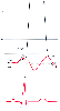 Figure 32-49
Relationship between left atrial pressure (LAP) and left
ventricular end-diastolic pressure (LVEDP). LVEDP is measured at the Z-point on
the left ventricular pressure (LVP) trace at the time of the electrocardiographic
R wave. Mean LAP (9 mm Hg) underestimates LVEDP (15 mm Hg), but the LAP a wave pressure
peak closely estimates LVEDP.[514]
(Redrawn
from Mark JB: Atlas of Cardiovascular Monitoring. New York, Churchill Livingstone,
1998, Fig. 6-1).
Figure 32-49
Relationship between left atrial pressure (LAP) and left
ventricular end-diastolic pressure (LVEDP). LVEDP is measured at the Z-point on
the left ventricular pressure (LVP) trace at the time of the electrocardiographic
R wave. Mean LAP (9 mm Hg) underestimates LVEDP (15 mm Hg), but the LAP a wave pressure
peak closely estimates LVEDP.[514]
(Redrawn
from Mark JB: Atlas of Cardiovascular Monitoring. New York, Churchill Livingstone,
1998, Fig. 6-1).
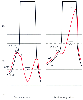 Figure 32-50
Relationship between left atrial pressure (LAP) and left
ventricular end-diastolic pressure (LVEDP) in aortic stenosis and mitral regurgitation.
LVEDP is elevated in both conditions (25 mm Hg), but mean LAP (
Figure 32-50
Relationship between left atrial pressure (LAP) and left
ventricular end-diastolic pressure (LVEDP) in aortic stenosis and mitral regurgitation.
LVEDP is elevated in both conditions (25 mm Hg), but mean LAP (![]() Ā
Ā![]() )
remains low in aortic stenosis (15 mm Hg) but high in mitral regurgitation (30 mm
Hg) because of the regurgitant c-v wave inscribed during systole. LVP, left ventricular
pressure. (Redrawn from Mark JB: Atlas of Cardiovascular Monitoring. New
York, Churchill Livingstone, 1998, Fig. 6-2).
)
remains low in aortic stenosis (15 mm Hg) but high in mitral regurgitation (30 mm
Hg) because of the regurgitant c-v wave inscribed during systole. LVP, left ventricular
pressure. (Redrawn from Mark JB: Atlas of Cardiovascular Monitoring. New
York, Churchill Livingstone, 1998, Fig. 6-2).
| Condition | Site of Discrepancy | Cause of Discrepancy |
|---|---|---|
| Diastolic dysfunction | Mean LAP < LVEDP | Increased end-diastolic a wave |
| Aortic regurgitation | LAP a wave < LVEDP | Mitral valve closure before end-diastole |
| Pulmonic regurgitation | PADP < LVEDP | Bidirectional runoff for pulmonary artery flow |
| Right bundle branch block | PADP < LVEDP | Delayed pulmonic valve opening |
| Postpneumonectomy | PAWP < LAP or LVEDP | Obstruction of pulmonary blood flow |
| LAP, left atrial pressure; LVEDP, left ventricular end-diastolic pressure; PADP, pulmonary artery diastolic pressure; PAWP, pulmonary artery wedge pressure. | ||
| Modified from Mark JB: Predicting left ventricular end-diastolic pressure. In Mark JB (ed): Atlas of Cardiovascular Monitoring. New York, Churchill Livingstone, 1998, p 59. | ||
When a PAC is used to estimate left ventricular filling pressure, in a number of situations this pressure is either underestimated ( Table 32-12 ) or overestimated ( Table 32-13 ) by the pulmonary artery diastolic or wedge pressure measurements. [513] Left ventricular diastolic dysfunction leading to decreased ventricular compliance is the most common cause of underestimation of left ventricular end-diastolic pressure.[515] The atrial contribution to left ventricular end-diastolic volume and pressure is normally less than 20%, but with diastolic dysfunction, this atrial contribution may approach 50%.[474] [477] Under these conditions, the wedge pressure a wave will be unusually prominent and will provide a close estimate of left ventricular end-diastolic pressure, whereas mean wedge pressure will underestimate left ventricular filling (see Fig. 32-42 ).[425] In patients with aortic regurgitation, abnormal diastolic left ventricular filling occurs from the aorta as soon as left ventricular pressure falls below aortic pressure and continues after left atrial contraction and closure of the mitral valve. Left ventricular diastolic pressure continues to rise until end-diastolic pressure is reached at the onset of mechanical systole, which reestablishes antegrade flow from the left ventricle to the aorta. Because mitral valve closure occurs before end-diastole, mean PAWP and the wedge pressure a wave both underestimate left ventricular end-diastolic pressure. [425]
In the presence of pulmonic regurgitation, diastolic flow from the proximal portion of the pulmonary artery becomes bidirectional, antegrade toward the left atrium and retrograde into the right ventricle. When right ventricular diastolic pressure is lower than left atrial pressure, pulmonary artery diastolic flow seeks the lower-pressure pathway toward the right ventricle, and pulmonary artery diastolic pressure will underestimate PAWP, left atrial pressure, and left ventricular end-diastolic pressure.[513]
When PVR is normal, there is no pressure gradient across the pulmonary vascular bed at the end of diastole, and pulmonary artery diastolic pressure equilibrates with the downstream left atrial pressure. However, when right ventricular systole is delayed because of right bundle branch block, PAP continues to fall with the x or
A final condition in which PAC measurements underestimate left ventricular end-diastolic pressure is different from the pathophysiologic states just noted because in this condition, the process of measuring wedge pressure changes pulmonary blood flow and thereby influences left heart filling. Under normal conditions, inflation of the PAC balloon to measure wedge pressure interrupts antegrade blood flow through a small section of the lung and has no measurable effect on total pulmonary blood flow or left heart filling. However, after pneumonectomy and possibly other conditions that markedly decrease the pulmonary vascular bed, balloon inflation may occlude a significant portion of the remaining pulmonary vascular cross-sectional area. Mechanical obstruction of pulmonary blood flow results and causes increased CVP and decreased left atrial pressure, cardiac output, and systemic blood pressure.[517] [518] Therefore, because PAC balloon inflation and measurement of wedge pressure lead to obstruction of pulmonary blood flow, the measured wedge pressure underestimates the left atrial and left ventricular end-diastolic pressures that existed before balloon inflation. This artifact should be suspected in the appropriate clinical setting, when a sudden reduction in systemic arterial pressure occurs coincident with PAC balloon inflation.
Respiratory influences on the pulmonary artery and wedge pressure
traces are the most common cause of overestimation of left ventricular end-diastolic
pressure ( Table 32-13
).
In this regard, mechanical ventilation with PEEP
poses a particular problem because it may create zone 1 or zone 2 lung conditions
and cause PAWP to be influenced by alveolar pressure.[519]
Formulas have been derived to adjust for the effects of PEEP on wedge pressure measurement.
In general, mechanical ventilation with PEEP increases wedge pressure by an amount
less than half the value of PEEP applied (i.e., 10 cm H2
O PEEP will raise
| Condition | Site of Discrepancy | Cause of Discrepancy |
|---|---|---|
| Positive end-expiratory pressure | Mean PAWP > mean LAP | Creation of lung zone 1 or 2 or pericardial pressure changes |
| Pulmonary arterial hypertension | PADP > mean PAWP | Increased pulmonary vascular resistance |
| Pulmonary veno-occlusive disease | Mean PAWP > mean LAP | Obstruction to flow in large pulmonary veins |
| Mitral stenosis | Mean LAP > LVEDP | Obstruction to flow across the mitral valve |
| Mitral regurgitation | Mean LAP > LVEDP | Retrograde systolic v wave raises mean atrial pressure |
| Ventricular septal defect | Mean LAP > LVEDP | Antegrade systolic v wave raises mean atrial pressure |
| Tachycardia | PADP > mean LAP > LVEDP | Short diastole creates pulmonary vascular and mitral valve gradients |
| LAP, left atrial pressure; LVEDP, left ventricular end-diastolic pressure; PADP, pulmonary artery diastolic pressure; PAWP, pulmonary artery wedge pressure. | ||
| Modified from Mark JB: Predicting left ventricular end-diastolic pressure. In Mark JB (ed): Atlas of Cardiovascular Monitoring. New York, Churchill Livingstone, 1998, p 59. | ||
When the pulmonary vasculature is normal, pulmonary blood flow ceases at end-diastole, and pulmonary artery diastolic pressure equals left atrial pressure.[524] However, if PVR increases, equilibration of PAP with downstream pressure does not occur. Under these conditions, the pulmonary vascular bed begins to resemble the systemic vasculature, where a large pressure gradient between systemic arterial pressure and right atrial pressure persists at end-diastole.
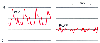 Figure 32-51
Pulmonary hypertension. The increased gradient across
the pulmonary vasculature causes pulmonary artery diastolic pressure to exceed pulmonary
artery wedge pressure (PAWP). PAP, pulmonary artery pressure. (Redrawn
from Mark JB: Atlas of Cardiovascular Monitoring. New York, Churchill Livingstone,
1998, Fig. 6-11.)
Figure 32-51
Pulmonary hypertension. The increased gradient across
the pulmonary vasculature causes pulmonary artery diastolic pressure to exceed pulmonary
artery wedge pressure (PAWP). PAP, pulmonary artery pressure. (Redrawn
from Mark JB: Atlas of Cardiovascular Monitoring. New York, Churchill Livingstone,
1998, Fig. 6-11.)
In contrast to the precapillary pulmonary vasoconstriction that exists in patients with pulmonary arterial hypertension, postcapillary obstruction to flow in the pulmonary veins may occur in the rare condition pulmonary veno-occlusive disease. Patients with this condition have normal left atrial pressure, but some disagreement exists regarding whether these patients have normal or elevated PAC-derived wedge pressure measurements.[120] [441] [525] [526] In part, these different observations may relate to whether the PAC has been wedged successfully because the wedge position may be difficult to obtain in these patients or, in some instances, the measured wedge pressure may be recorded from a totally occluded vascular channel. The most important factor, however, is whether the patient has predominantly small-vein occlusion or large-vein occlusion. Obstruction to flow in the small pulmonary veins narrows the static column of blood connecting the wedged PAC tip with the flowing column in the larger pulmonary veins. Given that the partial obstruction involves only the static column, wedge pressure will measure normal pulmonary venous and left atrial pressure because there is no pressure drop across this partially obstructed vascular segment in the absence of flow. Conversely, large-vein obstruction creates a pressure gradient across the large veins as blood flows in them toward the left atrium. In this instance, wedge pressure will detect an increased pulmonary venous pressure and overestimate left atrial and ventricular diastolic pressure. [120] [441] [525] [526] This model of pulmonary vein obstruction is consistent with the observations of Zidulka and Hakim, who measured wedge pressure in both large and small pulmonary arteries.[422] Other conditions may mimic the hemodynamic features of pulmonary veno-occlusive disease, including mediastinal fibrosis and intrathoracic or atrial tumors that obstruct pulmonary venous flow near the left atrium.
Mitral stenosis obstructs blood flow between the left atrium and left ventricle, and consequently, left atrial pressure will exceed left ventricular pressure throughout diastole. Furthermore, all upstream pressures recorded by the PAC will overestimate left ventricular end-diastolic pressure as a result of the valvular obstruction.[527] Because the pressure gradient across the mitral valve is directly related to the flow across it, pulmonary artery diastolic and wedge pressures will overestimate left ventricular filling pressures even more when transmitral flow is increased by tachycardia or elevated cardiac output.
Left atrial pressure is increased in mitral regurgitation because of the abnormal leakage of blood across the incompetent valve during systole, and left ventricular end-diastolic pressure is better approximated by measuring wedge pressure before onset of the regurgitant c-v wave (see Fig. 32-38 and Fig. 32-39 ). [458] In contrast to patients with mitral stenosis, in whom left atrial pressure exceeds left ventricular pressure throughout diastole, a patient with mitral regurgitation has appropriate diastolic equilibration of atrial and ventricular pressures. The problem in mitral regurgitation is choosing the appropriate end-diastolic, pre-v wave pressure to use as an estimate of left ventricular end-diastolic pressure. Mean PAWP will overestimate left ventricular end-diastolic pressure in any patient whose wedge pressure displays tall systolic v waves, including those with ventricular septal defect and left ventricular failure.[456] [457]
When tachycardia develops, the duration of diastole is shortened, and there is less time for egress of blood from the pulmonary vasculature to the left atrium and from the left atrium to the left ventricle. At both the mitral valve and pulmonary vascular levels, pressure gradients develop as the duration of diastole progressively decreases during tachycardia.[424] [514] [528] Consequently, pulmonary artery diastolic pressure overestimates mean PAWP, which in turn overestimates left ventricular end-diastolic pressure. Patients in atrial fibrillation will have more complete equilibration of pulmonary artery diastolic pressure and left ventricular end-diastolic pressure during longer R-R intervals. Consequently, these beats should be chosen to provide the best estimate of left ventricular end-diastolic pressure and preload.
In summary, there are many circumstances in which pulmonary artery diastolic or wedge pressure may underestimate or overestimate left ventricular end-diastolic pressure and thereby provide a misleading estimate of ventricular filling or preload. Some conditions are relatively common, whereas others are extremely rare, and frequently, several conditions coexist, further complicating the measurement problem. For example, a patient with mitral stenosis resulting in pulmonary hypertension in whom tachycardia develops has three reasons for the PAC-derived data to be in error and lead to a pulmonary artery diastolic pressure that markedly overestimates left ventricular end-diastolic pressure. Major clinical errors would result if the high pulmonary artery diastolic pressure is presumed to indicate increased left ventricular preload in a patient with these conditions.
|
|
|
|
|
|
|
|
|
|
|
|
|