|
|
|
|
|
|
|
|
|
|
|
|
|
|
|
Oximeters are devices that use light absorbance measurements to determine the concentration of various species of hemoglobin. One of the first in vivo oximeters was a noninvasive monitor used in aviation research during World War II. This device transilluminated tissue (the earlobe) with the light of two wavelengths. One wavelength was sensitive to changes in oxyhemoglobin, and the other was not. In effect, the earlobe acted as a test tube containing the suspended hemoglobin. For a review of the development of pulse oximetry, the reader is referred to an article by Severinghaus and Astrup.[19]
Before specific engineering problems are discussed, we need to define what we wish to measure. Adult blood usually contains four species of hemoglobin: oxyhemoglobin (HbO2 ), reduced hemoglobin (Hb), methemoglobin (metHb), and carboxyhemoglobin (COHb). Each of these hemoglobin species has a different light absorption profile. Figure 30-34
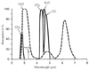 Figure 30-33
Absorption spectra of some gases and materials important
to anesthesia. Note that absorption is not constant over wavelengths. Therefore,
it is important to choose the proper wavelength to measure. In addition, when multiple
substances are present, it is still possible to measure their concentrations, provided
that enough wavelengths are available. (The solution becomes one of multiple equations
with multiple unknowns.) (From Gravenstein JS, Paulus DA, Hayes TJ: Capnography
in Clinical Practice. Boston, Butterworths, 1989.)
Figure 30-33
Absorption spectra of some gases and materials important
to anesthesia. Note that absorption is not constant over wavelengths. Therefore,
it is important to choose the proper wavelength to measure. In addition, when multiple
substances are present, it is still possible to measure their concentrations, provided
that enough wavelengths are available. (The solution becomes one of multiple equations
with multiple unknowns.) (From Gravenstein JS, Paulus DA, Hayes TJ: Capnography
in Clinical Practice. Boston, Butterworths, 1989.)
Measuring this quantity requires four wavelengths of light and
produces four simultaneous Beer-Lambert equations to solve for the four hemoglobin
species. Because metHb and COHb do not contribute to oxygen transport, functional
saturation is defined as the ratio of HbO2
to HbO2
plus reduced hemoglobin:
SaO2
= HbO2
/(HbO2
+ Hb)
Although functional saturation depends explicitly on only HbO2 and Hb, four light wavelengths are still required to measure it in the presence of significant concentrations of COHb and metHb.[20] [21] If the concentrations of metHb and COHb are both zero, O2 Hb% and SaO2 become identical.
The pulse oximeter performs substantial signal processing of optically transduced physiologic data. Although the principle governing pulse oximetry is straightforward, application of this principle to produce a clinically useful device involves significant engineering problems.[20] [22] The remainder of this section describes the physical and physiologic problems of pulse oximeter design and the engineering solutions to these problems. The discussion is divided into basic design and management of signal artifacts.
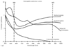 Figure 30-34
Hemoglobin extinction curves. Pulse oximetry uses the
wavelengths of 660 and 940 nm because they are available in solid-state emitters
(not all wavelengths are able to be emitted from diodes). Unfortunately, HbCO and
HbO2
absorb equally at 660 nm. Therefore, HbCO and HbO2
both
read as SaO2
to a conventional pulse oximeter.
In addition, Hbmet and reduced Hb share absorption at 660 nm and interfere with
correct SaO2
measurement. (Courtesy
of Susan Manson, Biox/Ohmeda, Boulder, Colorado, 1986.)
Figure 30-34
Hemoglobin extinction curves. Pulse oximetry uses the
wavelengths of 660 and 940 nm because they are available in solid-state emitters
(not all wavelengths are able to be emitted from diodes). Unfortunately, HbCO and
HbO2
absorb equally at 660 nm. Therefore, HbCO and HbO2
both
read as SaO2
to a conventional pulse oximeter.
In addition, Hbmet and reduced Hb share absorption at 660 nm and interfere with
correct SaO2
measurement. (Courtesy
of Susan Manson, Biox/Ohmeda, Boulder, Colorado, 1986.)
Noninvasive in vivo oximeters measure red and infrared light transmitted through and reflected by a tissue bed. Accurate estimation of SaO2 by this method entails several technical problems. First, there are many light absorbers in the transmitted light path other than arterial hemoglobin (e.g., skin, soft tissue, and venous and capillary blood). The pulse oximeter accounts for the effects of absorption of light by tissue and venous blood by assuming that only arterial blood pulsates. Figure 30-35 schematically illustrates the series of absorbers in a typical sample of living tissue. At the top of the figure is the AC component, which represents absorption of light by the pulsating arterial blood. The DC (baseline) component represents absorption of light by the tissue bed, including venous, capillary, and nonpulsatile arterial blood. Pulsatile expansion of the arteriolar bed increases the light path length (Equation 10 and Equation 11), thereby increasing absorbency. Pulse oximeters use only two wavelengths of light: 660 nm (red light) and 940 nm (near-infrared light). The pulse oximeter first determines the AC component of absorbance at each wavelength and then divides this value by the corresponding DC component to obtain a "pulse-added" absorbance, which is independent of the incident light intensity. The oximeter
 Figure 30-35
Pulse oximetry signals. A primary difficulty with pulse
oximetry is that the pulsatile signal is small in comparison to the total absorbance
of the ear or finger being examined. Pulsatile flow is needed to determine SaO2
.
(Adapted from Ohmeda Pulse Oximeter Model 3700 Service Manual.)
Figure 30-35
Pulse oximetry signals. A primary difficulty with pulse
oximetry is that the pulsatile signal is small in comparison to the total absorbance
of the ear or finger being examined. Pulsatile flow is needed to determine SaO2
.
(Adapted from Ohmeda Pulse Oximeter Model 3700 Service Manual.)
Finally, the value of R is related to the displayed saturation estimate SpO2 by a "look-up table" programmed into the oximeter's software. The tables used in all commercial pulse oximeters are based on experimental studies in healthy human volunteers. Although each manufacturer's exact calibration curve is proprietary, these curves are very similar. For instance, when the ratio of red-to-infrared pulse-added absorbance is 1.0, the displayed SpO2 is approximately 85%. This fact has clinical implications, which are discussed in the next section.
Probably the most difficult engineering problem in pulse oximetry is identification of the fluctuating absorbance pattern of arterial blood in a sea of electromagnetic and other artifacts. Artifacts have three major sources: ambient light, low perfusion (weak pulse, low AC-to-DC signal ratio), and motion (high AC-to-DC signal ratio). All these sources of artifacts produce a low signal-to-noise ratio.
The photodiodes used in the sensor to detect light cannot differentiate one wavelength of light from another. Therefore, the detector does not know whether the light received originates from the red light-emitting diode (LED), the infrared LED, or the room lights. This problem is solved in most pulse oximeters by alternating the red and infrared LED sources. The red LED is turned on first, and the photodiode detector produces a current resulting from the red LED plus the room lights. Next, the red LED is turned off and the infrared LED is turned on. The photodiode signal then represents the infrared LED plus the room lights. Finally, both LEDs are turned off, and the photodiode generates a signal from the room lights alone. This sequence is repeated hundreds of times per second. In this way, the oximeter attempts to eliminate light interference even in a quickly changing background of room light. [19] Some sources of fluctuating light can cause problems despite this clever design. Artifact from ambient light can be minimized simply by covering the sensor with an opaque shield.
Another engineering problem is that of a low AC-to-DC signal ratio, or low perfusion. When a small pulsatile absorbance signal is detected, the pulse oximeter amplifies the signal and estimates saturation from the ratio of the amplified absorbances. In this way, the pulse oximeter can estimate saturation values of SpO2 for a wide range of patients who generate different amplitudes for pulsatile absorbance. Unfortunately, as with a radio receiver, when a weak signal is amplified, the background noise, or "static," is also amplified. At the highest amplifications (which can be up to a million times), some pulse oximeters may analyze this noise signal and generate an SpO2 value from it. Because the noise is usually equal in the red and the infrared signals, the ratio of the two is often near unity (1.0), which yields a displayed saturation of approximately 85%. This problem could be demonstrated in early pulse oximeters by placing a piece of paper in the sensor between the photodiode and the LED. Many early models amplified the background noise while searching for a pulse until they eventually displayed a pulse and saturation value derived from the noise. To prevent this type of artifact, manufacturers have now incorporated minimum values for the signal-to-noise ratio, below which the device displays no value for SpO2 . Some oximeters also display a low-signal strength error message; in addition, many display a plethysmographic wave for visual identification of noise.
Patient motion (high AC-to-DC signal ratio) may be the most difficult artifact to eliminate. Engineers have tried several approaches to this problem, beginning with increasing the signal-averaging time. If the device averages its measurements over a longer period, the effect of an intermittent artifact is usually less. However, this longer averaging period also slows the response time to any acute change in SaO2 . Most pulse oximeters now allow the user to select one of several time-averaging modes. In addition, the designer can use sophisticated algorithms to identify and reject spurious signals.
One innovation aimed at reducing motion artifact is based on the premise that motion causes pulsations of venous blood within the tissue bed. Conventional pulse oximetry cannot distinguish these venous pulsations from those of arterial blood; hence, large errors or loss of signal can result. In the new signal-processing algorithm developed by Masimo, the oximeter actually computes a venous "noise reference signal," which is common to both light wavelengths. The noise reference is then subtracted from the total signal, and a "true" arterial signal is left. Tests in human volunteers as well as preliminary clinical studies indicate that this new technology represents an improvement in pulse oximeter performance in low signal-to-noise ratio situations.[23] [24]
Over the past decade, other in vivo oximeters have been developed to measure light reflected by living tissue in an attempt to determine tissue hemoglobin saturation in specific organs. Using reflected rather than transmitted light adds complexity because the path length of the light through the tissue may be tortuous and make calibration difficult. Nevertheless, a signal reflected from living tissue may produce useful information regarding the average saturation of the hemoglobin within the tissue illuminated by the reflectance oximeter.[25] For example, a cerebral oximeter may be able to measure mean brain hemoglobin saturation, which reflects the intracerebral balance between venous and arterial blood, as well as the oxygenation of both.
|
|
|
|
|
|
|
|
|
|
|
|
|