Aortic Diseases (also see Chapter
52
)
Aneurysms
Thoracic aortic aneurysms are much less common than abdominal
aortic aneurysms. Within the thorax, aortic aneurysms occur most frequently in the
ascending aorta, followed by the descending aorta. Aneurysms of the arch only are
uncommon. Thoracoabdominal aneurysms are aneurysms of the descending thoracic aorta
that extend into the abdominal aorta. Aneurysms of the ascending and descending
aorta tend to have different etiologies and natural histories and are managed differently.
Aneurysms of the ascending aorta most often result from cystic medial necrosis.
This disorder of smooth muscle and elastic fiber in the wall of the aorta is a frequent
finding in patients with connective tissue disorders such as Marfan's syndrome.
However, many patients with ascending aortic aneurysms have idiopathic cystic medial
necrosis of the aorta. Descending aortic aneurysms are most often associated with
atherosclerosis. Uncommon causes of thoracic aortic aneurysms include syphilis,
mycotic infections, aortic trauma, and confined aortic dissection.
Approximately 40% of patients with thoracic aortic aneurysms are
symptomatic at diagnosis. Patients typically have symptoms secondary to some vascular
consequence of the aneurysm or secondary to a local compressive/mass effect. Vascular
consequences can include aortic incompetence with symptoms of congestive heart failure
(CHF), coronary ischemia as a result of aneurysmal compromise of the coronary ostia
and proximal coronary arteries, and thromboembolic events. Rarely, thoracic aortic
aneurysms can rupture into the heart itself, especially the left atrium. Symptoms
may also be induced as a result of compression of local structures, such as the tracheobronchial
tree, the superior vena cava, or the esophagus.
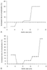 Figure 50-25
A, Influence of aortic
size on the cumulative lifetime incidence of natural complications of an aortic aneurysm.
On the y axis is plotted the incidence of natural
complications (rupture or dissection); on the x axis,
aortic size is plotted. This plot is for the ascending aorta. Note the hinge point
at 6 cm. B, The same plot for the descending aorta.
Note the hinge point at 7 cm.
Figure 50-25
A, Influence of aortic
size on the cumulative lifetime incidence of natural complications of an aortic aneurysm.
On the y axis is plotted the incidence of natural
complications (rupture or dissection); on the x axis,
aortic size is plotted. This plot is for the ascending aorta. Note the hinge point
at 6 cm. B, The same plot for the descending aorta.
Note the hinge point at 7 cm.
Patients with symptomatic thoracic aortic aneurysms are always
managed surgically. Gott and colleagues noted that nearly half of their marfanoid
patients with an aortic root greater than 6.5 cm had aortic dissection.[155]
Hence, they recommend prophylactic surgical repair in patients with an aneurysmal
size of even less than 6.5 cm. Other studies[156]
have examined complications in patients with ascending and descending thoracic aortic
aneurysms as a function of aneurysm size ( Fig.
50-25
) and have recommended criteria for prophylactic surgical intervention
for Marfan- and non-Marfan-related aneurysms at each location ( Table
50-6
).[156]
Dissections
Aortic dissections are not common, but they are frequently catastrophic.
The mechanisms underlying the development of aortic dissection are not necessarily
agreed on. Most experts believe that aortic dissections begin with an intimal tear
that results in the development of an intimal flap. They are observed most frequently
(65%) in the ascending aorta. However, autopsy studies do not always
identify an intimal tear, and it has been suggested that aortic dissections begin
in the media of the vessel wall with rupture of the vasa vasorum and subsequent rupture
into the vessel lumen ( Fig. 50-26
).
[157]
In patients with connective tissue disorders
(e.g., Marfan's syndrome), cystic medial degeneration causes weakness of the vessel
wall and predisposes to dissection. Patients without connective tissue disorders
lack the classic histologic features of medial degeneration. However, the degree
of medial degeneration in these patients far exceeds that observed in an appropriately
age-matched population. Advanced age and hypertension are the two most important
factors associated with medial degeneration and aortic dissection in these patients.
Other clinical conditions are also associated with dissection, including bicuspid
aortic valves, coarctation, pregnancy, blunt trauma, and postaortotomy.
Aortic dissections are classified by location ( Table
50-7
, Fig. 50-27
)
[157]
and duration. The use of more than one classification
to characterize location is potentially confusing, but can be simplified in that
ascending dissections (DeBakey types I and II, Stanford type A) require emergent
surgical intervention whereas descending dissections (DeBakey type III, Stanford
type B) are initially treated medically by controlling blood pressure. Acute dissection
describes those less than 2 weeks. After 2 weeks, the mortality secondary to dissection
levels off, thus rendering the management of chronic dissections very different from
that of acute dissections.
Patients with dissections most often have severe pain (sometimes
described by patients as ripping or tearing) that may migrate with extension of the
dissection. Ascending dissection is associated with anterior chest pain, whereas
involvement of the descending aorta is associated with back pain. Patients may also
have acute CHF (secondary to acute aortic incompetence), syncope, cerebral vascular
accidents, or cardiac arrest.