|
|
|
|
|
|
|
|
|
|
|
|
|
|
|
To perform spinal anesthesia, pertinent anatomy must be constantly kept in mind while inserting the spinal needle. It is often helpful for the technique to be broken down into a series of steps (i.e., the four Ps): preparation, position, projection, and puncture.
|
|
Dose (mg) * | Duration (min) | ||
|---|---|---|---|---|
| Local Anesthetic Mixture | To T10 | To T4 | Plain | Epinephrine, 0.2 mg |
| Lidocaine (5% in 7.5% dextrose) | 50–60 | 75–100 | 60 | 75–100 |
| Tetracaine (0.5% in 5% dextrose) | 6–8 | 10–16 | 70–90 | 100–150 |
| Bupivacaine (0.75% in 8.5% dextrose) | 8–10 | 12–20 | 90–120 | 100–150 |
| Ropivacaine (0.5% in dextrose) | 12–18 | 18–25 | 80–110 | — |
| Levobupivacaine | 8–10 | 12–20 | 90–120 | 100–150 |
Preparation of the equipment and drugs is essential for subarachnoid injection. When choosing a drug for subarachnoid injection, the duration of block should be matched to the surgical procedure and to patient variables ( Table 43-2 ). It may be more time efficient to use a hypobaric solution in a patient in the prone-jackknife position, rather than turning the patient after a hyperbaric solution has been allowed time to take effect. Is the patient planning to go home after an outpatient procedure, dictating a shorter-acting drug? Will a prolonged period of lower extremity analgesia, such as that obtained from adding an opioid to the spinal drugs, be advantageous to a patient undergoing knee reconstruction and immediate postoperative use of a range-of-motion machine? These are only a few of the many clinical permutations that dictate familiarity with the combinations of drugs available for subarachnoid use.
When choosing equipment for spinal anesthesia, the initial choice involves reusable or disposable equipment. Most anesthesiologists are forced to accept disposable spinal trays, which may seem limiting, but suppliers are willing to customize trays to meet professional preferences. Despite the emphasis on disposable trays, there is no indication that these trays shift the spinal anesthesia risk-benefit equation in the patient's favor.[59]
Spinal needles fall into two main categories: those that cut the dura and those designed to spread dural fibers. The former include the traditional disposable spinal needle, the Quincke-Babcock needle, and the latter contain the Whitacre and Sprotte needles ( Fig. 43-8 ). If a continuous spinal technique is chosen, the use of a Tuohy or other thin-walled needle can facilitate passage of the catheter. The use of small needles reduces the incidence of postdural puncture headache, whereas the use of larger needles improves the tactile sense of needle placement. Multiple punctures probably increase the incidence of headaches. If use of a smaller needle increases the number of punctures, the difference between small and large needles in producing headaches may be reduced. There is also a decrease in the incidence of postdural puncture headache when a conical-tipped needle is used, even when needle sizes are comparable.[60] Nevertheless, as facility with spinal anesthesia increases, the use of a smaller, similarly tipped needle can decrease headache incidence if the number of dural punctures does not increase.
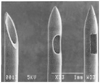 Figure 43-8
Spinal needle tip designs: Quincke (left),
Sprotte (middle), and Whitacare (right).
Scanning electron micrograph. (Adapted from Puolakka R, Andersson LC, Rosenberg
PH: Microscopic analysis of three different spinal needle tips after experimental
subarachnoid puncture. Reg Anesth Pain Med 25:163–169, 2000.)
Figure 43-8
Spinal needle tip designs: Quincke (left),
Sprotte (middle), and Whitacare (right).
Scanning electron micrograph. (Adapted from Puolakka R, Andersson LC, Rosenberg
PH: Microscopic analysis of three different spinal needle tips after experimental
subarachnoid puncture. Reg Anesth Pain Med 25:163–169, 2000.)
Positioning (see Chapter 28 ) is frequently the most poorly managed part of spinal technique for at least two reasons. First, the assistant often does not understand the rationale for positioning a patient, and second, patients are often inadequately or excessively sedated, making cooperation poor. The three primary methods of patient positioning include lateral decubitus, sitting, and prone, each with advantages in specific situations.
The lateral decubitus position is the most commonly used because it allows easier administration of more sedation and is less dependent on a well-trained assistant than with the sitting position. Patients are placed with the back parallel to the edge of the operating table nearest the anesthesiologist, with thighs flexed on the abdomen and the neck flexed to allow the forehead to be as close as possible to the knees. The assistant may be invaluable during this positioning by encouraging and assisting a patient in assuming the ideal lateral decubitus position ( Fig. 43-9 ). The patient should be positioned so that spread of hypobaric, isobaric, or hyperbaric solution to the operative site is optimized.
The sitting position should be chosen when low lumbar and sacral levels of sensory anesthesia are adequate for the surgical procedure, such as perineal and urologic operations, or when obesity or scoliosis makes identification of midline anatomy difficult in the lateral position. When placing patients in this position, a stool can be provided as a footrest, and a pillow is placed in the lap. The assistant then maintains the patient in a vertical plane while flexing the patient's neck and arms over the pillow to open up the lumbar vertebral space ( Fig. 43-10 ). If the reason for choosing the sitting position is to keep the sensory level low, the patient should be maintained sitting for 5 minutes; if the choice was made because of obesity or scoliosis and a higher sensory level is needed, the patient should be put supine immediately after subarachnoid injection, with the table manipulated appropriately. The most common error made in placing patients in this position is allowing the patient to slump, which loses the advantage of improved midline identification.
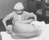 Figure 43-9
Lateral decubitus positioning for neuraxial block. The
assistant can help the patient assume the ideal position of "forehead to knees."
Figure 43-9
Lateral decubitus positioning for neuraxial block. The
assistant can help the patient assume the ideal position of "forehead to knees."
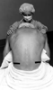 Figure 43-10
Sitting position for neuraxial block. The assistant
provides the patient with a foot rest (stool) and a pillow and prevents the patient
from slumping to either side.
Figure 43-10
Sitting position for neuraxial block. The assistant
provides the patient with a foot rest (stool) and a pillow and prevents the patient
from slumping to either side.
The prone position should be chosen when the patient is to be maintained in that position (often with jackknife modification) during the surgical procedure. This position is often appropriate for rectal, perineal, or lumbar procedures. An
 Figure 43-11
Spinal needle insertion. A,
The palpating fingers are "rolled" in a side-to-side and a cephalad-to-caudad direction
to identify interspinous space. B, During needle
insertion, the needle should be stabilized in a tripod fashion while placed in the
hand, similar to a dart being thrown.
Figure 43-11
Spinal needle insertion. A,
The palpating fingers are "rolled" in a side-to-side and a cephalad-to-caudad direction
to identify interspinous space. B, During needle
insertion, the needle should be stabilized in a tripod fashion while placed in the
hand, similar to a dart being thrown.
After the equipment, local anesthetics and additives, and the patient have been properly prepared, the midline or paramedian spinal puncture can be performed. The midline approach relies on the ability of patients and assistants to minimize lumbar lordosis and allow access to the subarachnoid space between adjacent spinous processes, usually at the L2–3, L3–4, or sometimes the L4–5 space. The palpating fingers (usually index and third fingers) should identify the interspinous area by identifying the caudad extent of the more cephalad spine and the midline by rolling the fingers medial to lateral ( Fig. 43-11 ). A subcutaneous skin wheal is developed overlying this space, and the introducer is inserted into the substance of the interspinous ligament. The introducer is grasped with the palpating fingers and steadied while the other hand is used to hold the spinal needle like a dart, and the fifth finger is used as a tripod against the patient's back to prevent patient movement producing unintentional insertion to a level deeper than intended. The needle, with bevel parallel to longitudinal dural fibers, is advanced slowly to heighten the sense of tissue planes traversed and to prevent skewing of nerve roots until the characteristic change in resistance is noted as the needle passes
After CSF is freely obtained, the dorsum of the anesthesiologist's nondominant hand steadies the spinal needle against the patient's back while the syringe containing the therapeutic dose is attached to the needle. CSF is again freely aspirated into the syringe, and the anesthetic dose is injected at a rate of approximately 0.2 mL/sec. After completion of the injection, 0.2 mL of CSF is aspirated into the syringe and reinjected subarachnoid to reconfirm location and to clear the needle of the remaining local anesthetic. The patient and operating table should then be placed in the position appropriate for the surgical procedure and drugs chosen.
The midline approach is the technique of first choice because it requires anatomic projection in only two planes
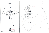 Figure 43-12
Vertebral anatomy of the midline and paramedian approaches
to centroneuraxis blocks. The midline approach highlighted in the inset
requires anatomic projection in only two planes: sagittal and horizontal. The paramedian
approach shown in the inset and in the posterior
view requires an additional oblique plane to be considered, although the technique
may be easier in patients who are unable to cooperate in minimizing their lumbar
lordosis. The paramedian needle insertion is made 1 cm lateral and 1 cm caudad to
the caudad edge of the more superior vertebral spinous process. The paramedian needle
is inserted approximately 15 degrees off the sagittal plane, as shown in the inset.
(Courtesy of the Mayo Foundation, Rochester, MN.)
Figure 43-12
Vertebral anatomy of the midline and paramedian approaches
to centroneuraxis blocks. The midline approach highlighted in the inset
requires anatomic projection in only two planes: sagittal and horizontal. The paramedian
approach shown in the inset and in the posterior
view requires an additional oblique plane to be considered, although the technique
may be easier in patients who are unable to cooperate in minimizing their lumbar
lordosis. The paramedian needle insertion is made 1 cm lateral and 1 cm caudad to
the caudad edge of the more superior vertebral spinous process. The paramedian needle
is inserted approximately 15 degrees off the sagittal plane, as shown in the inset.
(Courtesy of the Mayo Foundation, Rochester, MN.)
The paramedian approach exploits the larger "subarachnoid target" that exists if a needle is inserted slightly lateral to the midline (see Fig. 43-12 ). The most common error made in using the paramedian technique is that the needle entry site is placed too far off the midline, making the vertebral lamina barriers to needle insertion. In the paramedian approach, the palpating fingers again identify the caudad edge of the cephalad spinous process, and a skin wheal is raised 1 cm lateral and 1 cm caudad to this point. A longer needle (e.g., 1.5 to 2 inches) is then used to infiltrate deeper tissues in a cephalomedial plane. The spinal introducer and needle are then inserted 10 to 15 degrees off the sagittal plane in a cephalomedial plane (see Fig. 43-12 ). Similar to the midline approach, the most common error is to angle the needle too far cephalad on initial insertion. Nevertheless, if the needle contacts bone, it is redirected slightly in a cephalad direction. If bone is again contacted, but at a deeper level, the slight cephalad angulation is continued because it is likely that the needle is being walked up the lamina. As in the midline approach, the characteristic feel of the ligaments and
A variation on the paramedian approach is the lumbosacral approach described by Taylor.[61] This technique is carried out at the L5-S1 interspace, the largest interlaminar interspace of the vertebral column. A 5-inch spinal needle is inserted in a cephalomedial direction through a skin wheal raised 1 cm medial and 1 cm caudad to the lowermost prominence of the posterosuperior iliac spine. If bone is encountered on first needle insertion, the needle is walked off the sacrum into subarachnoid space. After CSF is obtained, the steps are similar to those previously outlined ( Fig. 43-13 ).
If a continuous spinal anesthetic is prescribed, a needle with a lateral-faced opening typically is used to perform the lumbar puncture ( Fig. 43-14 ). A midline or paramedian approach may be used, with some authorities suggesting that use of the paramedian approach facilitates catheter insertion.[62] The catheter should be threaded 2 to 3 cm into the subarachnoid space, and then the needle is withdrawn over the catheter. Care must be taken to ensure that the catheter is not inserted more deeply into the subarachnoid space when the needle is withdrawn over the catheter. Catheter-over-the-needle devices are also available for use with continuous spinal anesthesia. They are promoted as minimizing the leak of CSF around the catheter.[63]
 Figure 43-13
Neuraxial anatomy of the Taylor approach to spinal anesthesia.
This is really a paramedian approach at the L5-S1 vertebral level. A skin mark
is made 1 cm caudad and 1 cm medial to one of the posterior superior iliac spines.
Through this skin mark, the needle is inserted in a cephalad and medial direction.
The needle is walked off the sacrum and into the largest interspinous space, the
L5-S1 intervertebral space. The posterior superior iliac spines are located immediately
anterior to the often present "skin dimples" found overlying the superior aspect
of the sacrum. (Courtesy of the Mayo Foundation, Rochester, MN.)
Figure 43-13
Neuraxial anatomy of the Taylor approach to spinal anesthesia.
This is really a paramedian approach at the L5-S1 vertebral level. A skin mark
is made 1 cm caudad and 1 cm medial to one of the posterior superior iliac spines.
Through this skin mark, the needle is inserted in a cephalad and medial direction.
The needle is walked off the sacrum and into the largest interspinous space, the
L5-S1 intervertebral space. The posterior superior iliac spines are located immediately
anterior to the often present "skin dimples" found overlying the superior aspect
of the sacrum. (Courtesy of the Mayo Foundation, Rochester, MN.)
|
|
|
|
|
|
|
|
|
|
|
|
|