|
|
|
|
|
|
|
|
|
|
|
|
|
|
|
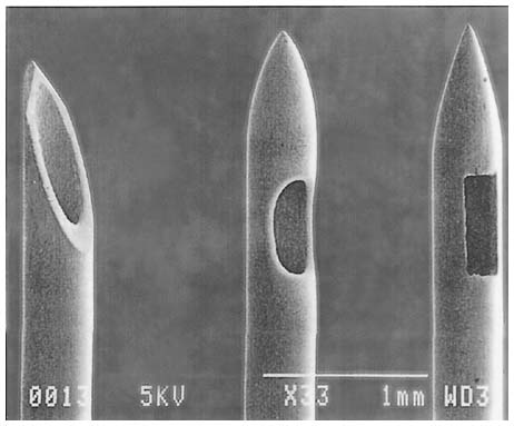
Figure 43-8
Spinal needle tip designs: Quincke (left),
Sprotte (middle), and Whitacare (right).
Scanning electron micrograph. (Adapted from Puolakka R, Andersson LC, Rosenberg
PH: Microscopic analysis of three different spinal needle tips after experimental
subarachnoid puncture. Reg Anesth Pain Med 25:163–169, 2000.)

|
|
|
|
|
|
|
|
|
|
|
|
|
|