Comprehensive Examination
In addition to the 8 cross sections described in the previous
section, 12 other cross sections are required to complete the comprehensive perioperative
TEE examination delineated in the 1999 ASE/SCA task force recommendations.[7]
When time permits or the diagnostic question or questions require, the comprehensive
examination can be completed in any order deemed most appropriate by the operator.
Shanewise and associates described an examination sequence based on anatomic structures,
and the reader is referred to this very detailed reference for one excellent approach.
[7]
However, when a single individual is responsible
for anesthetic care and TEE, the comprehensive examination is completed as time permits
after the basic examination has been recorded. When such is the case, the operator
can use 3 of the 8 basic cross sections as takeoff points for the other 12 cross
sections to make completion of the comprehensive examination a relatively easy sequence
to remember and quick to perform.
With the probe positioned for the AV SAX cross section, the other
six cross sections of the aorta are easily achieved. First, the probe is withdrawn
slowly 1 to 3 cm while keeping the aorta in the center of the video screen to view
the ascending aorta in SAX ( Fig. 33-8O
).
As the probe is withdrawn, the operator views progressively more superior SAX cross
sections of the aorta, beginning with the sinotubular junction, until the image is
lost because of interposition of the trachea between the esophagus and aorta. Next,
the multiplane angle is rotated forward to 100 to 120 degrees and advanced slowly
1 to 3 cm while keeping the aorta in the center of the video screen to view the ascending
aorta in LAX ( Fig. 33-8P
).
These two cross sections are ideal for assessment of pathology of the ascending
aorta, such as type I aortic dissection and aortic atheromas. However, the most
superior aspect of the aorta is rarely seen, including the takeoff of the innominate
artery, because of the aforementioned tracheal interposition. Next, the transducer
is returned to 0 degrees, the image depth decreased to 6 cm, and the probe turned
leftward past the cardiac structures to reveal the descending aorta in SAX ( Fig.
33-8Q
). The probe is withdrawn and advanced with the aorta maintained
in the center of the screen until the entire descending aorta has been examined in
SAX. This maneuver is repeated with the transducer at 90 degrees to examine the
descending aorta in LAX ( Fig. 33-8R
).
Next, the transducer is returned to 0 degrees and the probe withdrawn until the
distal aortic arch in LAX comes into view ( Fig.
33-8S
). Rotating the transducer to 90 degrees reveals the distal arch
in SAX ( Fig. 33-8T
). The
probe is turned leftward until the aorta just disappears from view and then turned
slowly rightward to identify the takeoff of the left subclavian artery (well seen
in most patients) and the left carotid artery (well seen in a minority of patients).
These cross sections of the descending aorta and distal aortic arch reliably reveal
dissections and atheromatous disease.
With the probe positioned for the ME four-chamber cross section,
the ME mitral commissural cross section is achieved easily by centering the coaptation
point of the mitral valve in the center of the video screen and rotating the multiplane
angle forward to about 60 degrees ( Fig.
33-8G
). This maneuver positions the ultrasound beam parallel to the closure
line of the mitral leaflets and completes the LAX examination of the mitral valve.
Although a detailed discussion of mitral leaflet structure and function is beyond
the scope of this chapter, Figure
33-10
summarizes an excellent approach to this challenging task.[35]
With the probe positioned for the TG mid SAX cross section, the
remaining five cross sections of the comprehensive
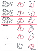 Figure 33-10
Systematic examination of the mitral valve. In this
examination, the mitral valve is viewed in multiple cross sections to delineate leaflet
anatomy. The "5-chamber" cross section is accomplished by withdrawing the probe
slightly from the standard 4-chamber cross section until the left ventricular outflow
track is in view. The center column shows the planes
of the different cross sections as viewed from directly above the base of the heart.
The 2-chamber "anterior," "mid," and "posterior" cross sections are variations of
the standard 2-chamber cross section and are accomplished by turning the probe from
the patient's right to left. "P1, P2, and P3" refer to the three scallops of the
posterior mitral leaflet, and "A1, A2, and A3" refer to the juxtaposed segments of
the anterior mitral leaflet. The right column shows
the leaflet segments seen in the corresponding cross section. (Redrawn from
Lambert AS, Miller JP, Foster E, et al: Improved evaluation of the location and
mechanism of mitral valve regurgitation with a systematic transesophageal echocardiography
examination. Anesth Analg 88:1205–1212, 1999.)
Figure 33-10
Systematic examination of the mitral valve. In this
examination, the mitral valve is viewed in multiple cross sections to delineate leaflet
anatomy. The "5-chamber" cross section is accomplished by withdrawing the probe
slightly from the standard 4-chamber cross section until the left ventricular outflow
track is in view. The center column shows the planes
of the different cross sections as viewed from directly above the base of the heart.
The 2-chamber "anterior," "mid," and "posterior" cross sections are variations of
the standard 2-chamber cross section and are accomplished by turning the probe from
the patient's right to left. "P1, P2, and P3" refer to the three scallops of the
posterior mitral leaflet, and "A1, A2, and A3" refer to the juxtaposed segments of
the anterior mitral leaflet. The right column shows
the leaflet segments seen in the corresponding cross section. (Redrawn from
Lambert AS, Miller JP, Foster E, et al: Improved evaluation of the location and
mechanism of mitral valve regurgitation with a systematic transesophageal echocardiography
examination. Anesth Analg 88:1205–1212, 1999.)
examination are easily achieved in most patients. First, the left ventricle is centered
in the video screen and the multiplane angle is rotated forward to 90 degrees to
reveal the TG two-chamber cross section ( Fig.
33-8E
). Although this cross section reveals the same structures as the
ME two-chamber cross section, the viewing angle is orthogonal to the former viewing
angle, and as a result, subvalvular structures are seen better. Next, the transducer
is rotated to 100 to 120 degrees to reveal the TG LAX cross section ( Fig.
33-8J
). Again, this cross section reveals the same structures as the ME
LAX, but the TG LAX view permits more parallel alignment of the ultrasound beam with
blood flow through the LV outflow
track and AV. Next, the TG RV inflow cross section is achieved by returning the
probe to the TG mid SAX position, turning it rightward until the right ventricle
is in the centered in the video screen, and then rotating the multiplane angle to
100 to 120 degrees to bring the RV apex into view ( Fig.
33-8N
). This cross section is ideal for viewing the RV inferior free wall.
Next, the TG basal SAX is achieved by returning the probe to the TG mid SAX, releasing
the flexion of the probe, withdrawing 1 to 2 cm, and then flexing it gently until
the orifice of the mitral valve is seen in SAX ( Fig.
33-8F
). This cross section can prove vital in determining the precise
location of mitral regurgitation.[35]
Finally,
the deep TG LAX cross section is achieved by returning the probe to the TG mid SAX
position, releasing the flexion of the probe, advancing it 6 to 8 cm into the stomach,
fully flexing it once in the stomach, gently withdrawing it until minimal resistance
is met at the gastroesophageal junction, and then slightly turning it leftward or
rightward to reveal the LV outflow track and AV ( Fig.
33-8K
). Usually, this cross section does not resolve structures as well
as the ME LAX does, but it does provide optimal beam alignment for Doppler interrogation
of the LV outflow track and AV. Often, this cross section is the most difficult
of the cross sections to achieve. If a few gentle attempts fail to accomplish it,
it should be abandoned.