Abbreviated Examination
Because of time constraints and relatively narrow diagnostic goals,
anesthesiologists often perform a more limited intraoperative examination than described
in the ASE/SCA's task force recommendation for a comprehensive TEE examination (see
the subsequent section).[7]
[34]
However, even when time is critical, the examination performed should allow at least
the basic applications of TEE as outlined in the 1996 guidelines for perioperative
TEE: detection of markedly abnormal ventricular filling or function, extensive myocardial
ischemia or infarction, large air embolism, severe valvular dysfunction, large cardiac
masses or thrombi, large pericardial effusions, and major lesions of the great vessels.
[6]
A minimum of eight different cross sections
drawn from the 20 cross sections delineated in the comprehensive examination are
required to meet these diagnostic goals. Four of the cross sections are imaged in
both two-dimensional and color Doppler to assess valvular function. The next paragraph
describes the probe manipulations required to achieve these cross sections. The
reader should review Figure 33-8
to understand the terms used in this description.
After the TEE probe is introduced safely into the esophagus, it
is advanced to the midesophageal (ME) level (28 to 32 cm measured at the upper incisors),
and the
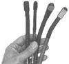 Figure 33-7
Four TEE probes compared. From left to right the probes
are single plane, pediatric single plane, multiplane, and biplane. The probe tips
are viewed in their maximum dimension. (From Cahalan MK: Intraoperative
Transesophageal Echocardiography. An Interactive Text and Atlas. New York, Churchill
Livingstone, 1997.)
Figure 33-7
Four TEE probes compared. From left to right the probes
are single plane, pediatric single plane, multiplane, and biplane. The probe tips
are viewed in their maximum dimension. (From Cahalan MK: Intraoperative
Transesophageal Echocardiography. An Interactive Text and Atlas. New York, Churchill
Livingstone, 1997.)
aortic valve (AV) is imaged in the short axis (SAX) by turning the probe, adjusting
its depth in the esophagus, and rotating the multiplane transducer to 25 to 45 degrees
until the three cusps of the valve are seen as approximately equal in size and shape
( Fig. 33-8H
). Image depth
is set at 10 to 12 cm as required to position the AV in the center of the video screen.
This cross section is ideal for detection of aortic stenosis. The videotape is
activated at this point and kept running throughout the rest of the examination.
Videotape is very inexpensive relative to the cost of a missed diagnosis. Next,
the probe is turned slightly to position the AV in the center of the video screen,
and the multiplane angle is then rotated forward to 110 to 130 degrees to bring the
long axis (LAX) of the AV in view ( Fig.
33-8I
). This cross section is best for detection of ascending aortic abnormalities,
including type I aortic dissection. Color Doppler is used for assessment of AV competence.
For detection of valvular stenosis and regurgitation, the maximum possible Nyquist
limit is used (ideally, above 50 cm/sec). Next, Doppler is discontinued and the
probe is turned rightward until the ME bicaval cross section comes into view ( Fig.
33-8L
). This cross section is usually seen best at a multiplane angle
between 90 and 110 degrees and is ideal for assessing caval abnormalities, compression
of the right atrium from anteriorly located masses or effusions, and compression
of the left atrium from posteriorly located masses or effusions. In addition, the
bicaval cross section may reveal collections of air located anteriorly in the left
or right atrium, as well as the structure of the interatrial septum, including the
foramen ovale. Next, the multiplane angle is rotated back to 60 to 80 degrees and
the probe is turned leftward just past the AV to bring the ME right ventricular (RV)
inflow
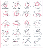 Figure 33-8
TEE cross sections in a comprehensive examination. Twenty
standard cross sections and their abbreviated names are depicted by the line drawings.
The text describes the probe manipulations required to produce each of the cross
sections. (Redrawn from Shanewise JS, Cheung AT, Aronson S, et al: ASE/SCA
guidelines for performing a comprehensive intraoperative multiplane transesophageal
echocardiography examination: Recommendations of the American Society of Echocardiography
Council for Intraoperative Echocardiography and the Society of Cardiovascular Anesthesiologists
Task Force for Certification in Perioperative Transesophageal Echocardiography.
Anesth Analg 89:870–884, 1999.)
Figure 33-8
TEE cross sections in a comprehensive examination. Twenty
standard cross sections and their abbreviated names are depicted by the line drawings.
The text describes the probe manipulations required to produce each of the cross
sections. (Redrawn from Shanewise JS, Cheung AT, Aronson S, et al: ASE/SCA
guidelines for performing a comprehensive intraoperative multiplane transesophageal
echocardiography examination: Recommendations of the American Society of Echocardiography
Council for Intraoperative Echocardiography and the Society of Cardiovascular Anesthesiologists
Task Force for Certification in Perioperative Transesophageal Echocardiography.
Anesth Analg 89:870–884, 1999.)
and outflow cross section into view ( Fig.
33-8M
). Usually, an image depth of 12 to 14 cm is required to position
the RV outflow track in the center of the video screen. This cross section reveals
the contractile function of the right ventricle, the outflow tract, and pulmonary
valve function with the application of color Doppler. Next, the transducer is rotated
back to 0 degrees and the probe is advanced 4 to 6 mm into the esophagus and gently
retroflexed until all four cardiac chambers are visualized (ME four-chamber cross
section) ( Fig. 33-8A
).
Often, rotating the transducer 10 to 15 degrees will enhance the view of the tricuspid
annulus. Generally, an image depth of 14 to 16 cm is required to include the LV
apex in the sector scan. In two-dimensional imaging, the free wall of the right
ventricle and the lateral and septal LV wall segments are evaluated for contractile
function. With color Doppler, both the mitral and tricuspid valves are assessed.
Stenotic and regurgitant lesions can be diagnosed accurately. During this assessment,
image depth is decreased to 10 to 12 cm to afford a magnified view of the valves
and maximization of the Nyquist limit (above 50 cm/sec). Next, color Doppler is
discontinued, the left ventricle is positioned in the center of the screen, and the
multiplane angle is rotated forward to 90 degrees to bring into view the ME two-chamber
cross section ( Fig. 33-8B
).
Image depth is returned to 14 to 16 cm. This cross section is best for revealing
the function of the basal and apical segments of the anterior and inferior LV walls,
as well as anterior and inferior pericardial collections. When air emboli collect
in the left ventricle, they can usually best be seen in this view as very echogenic
areas located along the anterior apical endocardial surface. The transducer is then
rotated forward to 135 degrees to reveal the ME LAX cross section that is best for
assessment of the anteroseptal and posterior wall segments for contractile LV function
( Fig. 33-8C
). Together,
the ME four-chamber, two-chamber, and LAX cross sections reveal all 16 segments of
the left ventricle ( Fig. 33-9
).
However, the next and last of the basic cross sections provides a second look at
the midventricular segments, as well as other benefits. To achieve this cross section,
the transducer is rotated back to 0 degrees, the left ventricle is centered in the
screen, and the probe
 Figure 33-9
Five TEE cross sections with myocardial segments identified.
A total of 16 myocardial segments are identified and named according to standards
adopted by the American Society of Echocardiography and the Society of Cardiovascular
Anesthesiologists. (Redrawn from Shanewise JS, Cheung AT, Aronson S. et
al: ASE/SCA guidelines for performing a comprehensive intraoperative multiplane
transesophageal echocardiography examination: Recommendations of the American Society
of Echocardiography Council for Intraoperative Echocardiography and the Society of
Cardiovascular Anesthesiologists Task Force for Certification in Perioperative Transesophageal
Echocardiography. Anesth Analg 89:870–884, 1999.)
Figure 33-9
Five TEE cross sections with myocardial segments identified.
A total of 16 myocardial segments are identified and named according to standards
adopted by the American Society of Echocardiography and the Society of Cardiovascular
Anesthesiologists. (Redrawn from Shanewise JS, Cheung AT, Aronson S. et
al: ASE/SCA guidelines for performing a comprehensive intraoperative multiplane
transesophageal echocardiography examination: Recommendations of the American Society
of Echocardiography Council for Intraoperative Echocardiography and the Society of
Cardiovascular Anesthesiologists Task Force for Certification in Perioperative Transesophageal
Echocardiography. Anesth Analg 89:870–884, 1999.)
is advanced 4 to 6 cm into the stomach. The probe is then flexed gently anteriorly
to reveal the transgastric (TG) SAX cross section ( Fig.
33-8D
). This cross section is ideal for monitoring LV filling and contractile
function. All major coronary arteries supplying the myocardium are viewed in this
cross section. Moreover, changes in preload cause greater changes in the LV SAX
dimension than the LV LAX dimension, and movement of the probe from this cross section
is readily apparent because the papillary muscles provide prominent landmarks. Since
this cross section is used to judge filling and ejection, image depth is consistently
set to 12 cm so that the size and function of the heart are judged easily relative
to previously examined hearts.