 |
 |
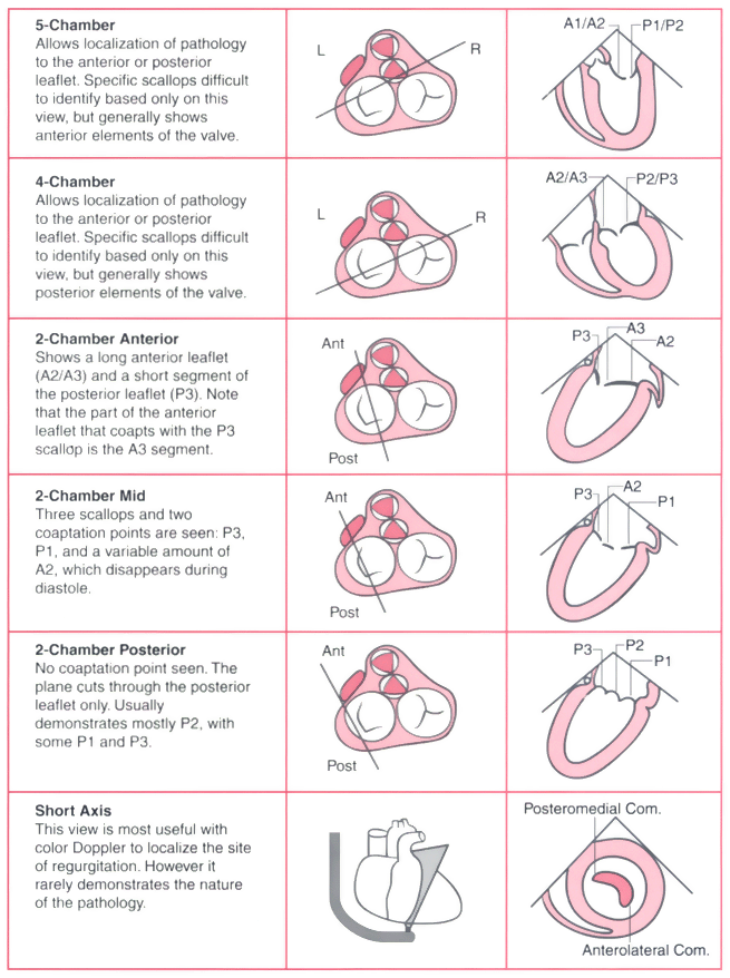
Figure 33-10
Systematic examination of the mitral valve. In this
examination, the mitral valve is viewed in multiple cross sections to delineate leaflet
anatomy. The "5-chamber" cross section is accomplished by withdrawing the probe
slightly from the standard 4-chamber cross section until the left ventricular outflow
track is in view. The center column shows the planes
of the different cross sections as viewed from directly above the base of the heart.
The 2-chamber "anterior," "mid," and "posterior" cross sections are variations of
the standard 2-chamber cross section and are accomplished by turning the probe from
the patient's right to left. "P1, P2, and P3" refer to the three scallops of the
posterior mitral leaflet, and "A1, A2, and A3" refer to the juxtaposed segments of
the anterior mitral leaflet. The right column shows
the leaflet segments seen in the corresponding cross section. (Redrawn from
Lambert AS, Miller JP, Foster E, et al: Improved evaluation of the location and
mechanism of mitral valve regurgitation with a systematic transesophageal echocardiography
examination. Anesth Analg 88:1205–1212, 1999.)

 |
