|
|
|
|
|
|
|
|
|
|
|
|
|
|
|
The first neuraxial block was performed 8 months after the demonstration in Heidelberg of the local anesthetic properties of cocaine (see Chapter 72 ). James Leonard Corning (1855–1923) was a neurologist who had learned
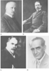 Figure 1-11
Pioneers of neuraxial block: A,
August K. G. Bier. B, Theodore Tuffier. C,
Rudolph Matas. D, Achille M. Dogliotti. (Courtesy
of the National Library of Medicine, Bethesda, MD.)
Figure 1-11
Pioneers of neuraxial block: A,
August K. G. Bier. B, Theodore Tuffier. C,
Rudolph Matas. D, Achille M. Dogliotti. (Courtesy
of the National Library of Medicine, Bethesda, MD.)
The consensus is that Corning produced an epidural injection of the drug[242] because 120 mg (60 mg initially and then 60 mg 8 minutes later) of intrathecal cocaine would be expected to produce a total spinal anesthetic or a block extending into the cervical dermatomes. It is not surprising that Corning's method of neuraxial block was not repeated because the technique of consistently injecting into the epidural space had not yet been described. If any investigators had attempted to repeat the Corning experiment with the dose he used, it might have ended in disaster and thereby delayed the development of neuraxial block by several years.
In Kiel, Germany, during the last decade of the 19th century, preparations were being made for the next major advance in anesthetic practice. At the Kiel University Medical School, Friederich von Esmarch (1823–1908) was the senior surgeon and August Bier one of the junior surgeons. Heinrich I. Quincke (1842–1922) was the leading internist in Kiel and had already contributed several
On August 15, 1898, August Bier and his assistant August Hildebrandt (1868–1854) used the Quincke method of entering the intrathecal space and injected between 5 and 15 mg of cocaine to produce spinal anesthesia in six cases for operations on the lower part of the body. They also reported the results of spinal anesthesia given to each other in what has become one of the classic clinical papers in the medical literature.[245] Bier thought it would not replace general anesthesia because of the severity of side effects such as nausea, vomiting, dizziness, and headache. He proposed that these undesirable side effects were caused by escape of cerebrospinal fluid from the dural sac. The method whereby Bier arrived at the correct intrathecal dose of cocaine on the first attempt remains a mystery.
After Bier's report, interest in spinal anesthesia spread rapidly. J. B. Seldowitsch[246] successfully provided spinal anesthesia in St. Petersburg on May 11, 1899; Frederick Dudley Tait (1862–1918) and Guido Caglieri[247] (1871–1951) in San Francisco on October 26, 1899; Theodore Tuffier[248] (see Fig. 1-11B ) in Paris on November 9, 1899; and Rudolph Matas[249] (1860–1957) (see Fig. 1-11C ) in New Orleans on November 10, 1899. By one report, more than 1000 manuscripts relating to spinal anesthesia had been published within 2 years of the original paper by Bier. [250]
Not all researchers agreed on the technique and indications for spinal anesthesia. Tait and Caglieri suggested the use of cervical intrathecal injections for operating on the upper extremities. A. W. Morton[251] reported success with total spinal anesthesia after lumbar puncture for operations on all parts of the body. Thomas Jonnesco[252] reported no adverse effects from 398 spinal anesthetics administered between the spines of the thoracic and lumbar levels using a novocaine and strychnine mixture. Jonnesco called the method general spinal anesthesia. Remarkably, in his series there were 14 operations on the skull, 45 on the face, and 25 on the neck. In 1909, Bier[253] claimed that to be successful with spinal anesthesia, the anesthetist should inject the solution only at body temperature and that tropacocaine was preferable to cocaine.
The early reports of cocaine spinal anesthesia mentioned that after injection, patients often became restless and excitable, often exhibiting a significant rise in body temperature. One of the first physicians to specialize in anesthesia, S. Ormond Goldan, maintained accurate anesthesia records from several cases of spinal anesthesia with cocaine.[254] His records reveal a typical increase in heart rate, pupil size, and body temperature after cocaine spinal anesthesia. Matas reasoned that these effects were secondary to an action of cocaine on the central nervous system. He learned that mixing 1.5 mg of morphine with cocaine was useful in mitigating these symptoms. In his 1900 report on spinal anesthesia, he regarded a mixture of cocaine and morphine as his standard agent. This report by Matas appears to be among the first attempts to use spinal opioids to enhance neuraxial analgesia.[249] The Japanese anesthesiologist Otojiro Kitagawa[255] (1864–1922) used intrathecal morphine (10 mg) in the same year to treat the chronic painful conditions of two patients.
It is not surprising that serious complications from the spinal technique were soon observed. F. Gumprecht[256] reported 15 cases of sudden death from lumbar puncture in 1900. Several investigators observed respiratory arrest after high spinal injections. After the introduction of routine blood pressure measurements by Cushing[257] in 1903, it was observed that severe hypotension could occur after spinal anesthesia. [258]
The scientific study of spinal anesthesia began within a few years after its introduction. Investigations were undertaken by Arthur E. Barker[259] (1850–1916) to determine the factors involved in spread of the local anesthetic within the subarachnoid space. Barker advised meticulous sterile technique and introduced the use of dextrose to produce hyperbaric solutions. His emphasis on gravity as an essential determinant of local anesthetic spread remains an important facet of the spinal technique today.[260] Several researchers reported the dangers of total spinal anesthesia. Gaston Labat[62] and George P. Pitkin[261] contributed clinical observations that improved the safety of spinal anesthesia.
A widely publicized malpractice trial in 1953 had a negative impact on the use of spinal anesthesia.[262] Albert Woolley and Cecil Roe were healthy subjects who received dibucaine spinal anesthetics on the same day at the Chesterfield Royal Hospital in England. Both patients developed permanent painful spastic paraparesis. Although the cause of paresis was inconclusive, it was thought that the injuries were caused by contamination of the spinal solution by phenol, in which the dibucaine ampules had been immersed for sterilization. The Wooley and Roe case was followed by other reports of paralysis after spinal anesthesia. [263] However, in 1954, a reassuring study of 10,098 spinal anesthetics with only 71 minor neuropathies, most unrelated to the block itself, was published in a widely circulated medical journal.[264] Spinal anesthesia then reemerged as a safe anesthetic method provided that attention was directed to meticulous technique.
Consideration has been given to a syndrome characterized by transient paresthesias after lidocaine spinal anesthesia.[265] [266] However, with the introduction of disposable spinal kits and improved techniques, the spinal route of drug administration is now firmly established. Research is continuing on new drugs and methods of delivery. Reports of cauda equina syndrome after the introduction of lidocaine through spinal microcatheters[267] emphasize the importance of careful clinical observations when new methods of spinal delivery are introduced.
Post-spinal headache was an annoying problem for the first practitioners and their patients. The exact cause for this reaction was not agreed on for several years.
The use of small-diameter spinal needles has decreased the incidence of spinal headache after spinal anesthesia. However, inadvertent dural puncture with larger needles can sometimes occur during the placement of epidural catheters. An innovative treatment for headache after dural puncture, the epidural blood patch, was suggested by James B. Gormley[269] in 1960 and further described by Anthony J. DiGiovanni and Burdett S. Dunbar[270] in 1970. The blood patch has been reported to be successful in a high percentage of cases and has withstood the test of time as an effective treatment for this condition.
|
|
|
|
|
|
|
|
|
|
|
|
|