|
|
|
|
|
|
|
|
|
|
|
|
|
|
|
It was not obvious that cocaine would produce blockade of sensation if injected directly into peripheral nerves. In 1880, von Anrep had injected cocaine under the skin of his arm and discovered that it produced insensitivity, but this information did not attract attention. At least one Viennese surgeon, Anton Wölfler, first assistant to Theodore Billroth, had attempted hypodermic cocaine injections without producing analgesia and was convinced that it was effective only on the mucous membranes. The idea of injecting cocaine into nerve trunks is credited to William Halsted (1852–1922) and Alfred Hall, who began their injection experiments as early as 8 weeks after the Heidelberg announcement.
Halsted and Hall had studied in Vienna during 1879 and 1880, but it is unlikely that they met Koller during those years, because Koller did not finish his medical school training until 1882. During those years, Koller was working on a paper about the development of the mesoderm and had not developed his interest in cocaine. In 1884, Halsted was occasionally performing operations in the bedroom of his own house in New York City, and it was there that the two surgeons began their work on regional anesthesia. The first report of their success with injection appeared on December 6, 1884 in the New York Medical Journal in a letter written by Hall.[217] In this letter, Hall reports that they first injected 4% cocaine (15 mg) into the forearm and concluded that it blocked transmission in the cutaneous nerves because it provided analgesia below but not above the point of injection. They then injected 2 mL (80 mg) into the ulnar nerve at the elbow, producing block of the entire ulnar distribution distal to the point of injection. Additional blocks were then performed on the brachial plexus and the infraorbital nerves, inferior dental nerves, and the sciatic nerve, all for operative surgery.[218]
With these large doses, it is not surprising that constitutional symptoms developed. Hall described dizziness and nausea. Both Halsted and Hall became addicted to cocaine; Halsted lived with an occult cocaine or morphine addiction the rest of his life. Hall took a position at Columbia University in New York City, but he later moved to Santa Barbara, California, where he died in 1924.
Carl Schleich[219] (1859–1922) introduced infiltration local anesthesia in 1892 as an alternative to direct injection of nerve trunks. His method was to infiltrate cocaine in dilute concentrations (0.01% to 0.2%) directly into the subcutaneous tissues. James Leonard Corning[220] (1855–1923), a neurologist from New York, observed that placing a tourniquet on the limb could prolong the analgesic effect of infiltration analgesia, and he reasoned that the tourniquet prevented the blood from removing cocaine from its active site. Heinrich F. Braun[221] [222] (1862–1934) achieved the same prolonged effect of cocaine by adding epinephrine to the solution, producing a "chemical tourniquet." Braun became the pioneer of the new drug procaine,[223] introduced by Braun in 1905 as a less toxic drug than cocaine. Braun's textbook,[221] first published in 1907, was one of the first devoted to regional anesthesia and went through eight editions, with the last one published in 1933.
Although Halsted was the first to block the brachial plexus, he did not use a percutaneous technique. His method in 1884, and that used by George Crile 13 years later, was to surgically expose the roots and then inject each nerve directly. G. Hirschel[224] produced the first percutaneous brachial plexus block in 1911 through an axillary approach ( Fig. 1-10A ). The axillary brachial plexus block has been modified by several surgeons, including George Pitkin,[225] and R. H. de Jong,[226] and remains a popular technique today.
D. Kulenkampff introduced the supraclavicular brachial plexus block[227] a few months after Hirschel described the axillary approach (see Fig. 1-10B ). Kulenkampff injected his own plexus with 10 mL of procaine at the midclavicular position, lateral to the subclavian artery, obtaining complete anesthesia of the arm. Early reports indicated a high degree of success with this block, but other practitioners soon reported complications such as pneumothorax and mediastinal emphysema. Several modifications of the supraclavicular block have emerged in an effort to avoid pneumothorax. Infraclavicular approaches to the brachial plexus were described by L. Bazy and V. Pauchet[228] in 1917 and later popularized by P. Raj[229] in 1973.
In an attempt to approach the brachial plexus in the neck and thereby avoid pulmonary complications, M. Kappis[230] (1912) attempted to perform the block through a posterior paravertebral approach. Because of a high incidence of failures with the posterior approach, several investigators, including J. Etienne,[231] V. Pauchet, and G. Pitkin, used various anterior approaches to the brachial plexus in the neck. In 1970, Alon P. Winnie[232]
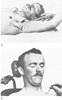 Figure 1-10
A, Hirschel performed
the first percutaneous axillary block with 20 mL of 2% procaine in 1911. He forced
a rubber ball under the pectoral muscles and fixed it with elastic bandages to prevent
rapid absorption of anesthetic solution, a maneuver that he later abandoned as unnecessary.
(From Hirschel G: Die Anasthesierung de Plexus Brachialis fur die Operationen
an der oberen Extremitat, Much Med Wochenschr 58:1555–1556, 1911.)
B, Kulenkampff approached the brachial plexus above
the clavicle, just lateral to the subclavian artery. The patients were positioned
in the sitting position for the block. An injection of 10 mL of 2% procaine with
epinephrine was given only after a paresthesia had been obtained. If the first rib
was contacted, the injection was made more medially. (From Kulenkampff D:
Anesthesia of the brachial plexus. Zentrabl Chr 38:1337–1340, 1911.)
Figure 1-10
A, Hirschel performed
the first percutaneous axillary block with 20 mL of 2% procaine in 1911. He forced
a rubber ball under the pectoral muscles and fixed it with elastic bandages to prevent
rapid absorption of anesthetic solution, a maneuver that he later abandoned as unnecessary.
(From Hirschel G: Die Anasthesierung de Plexus Brachialis fur die Operationen
an der oberen Extremitat, Much Med Wochenschr 58:1555–1556, 1911.)
B, Kulenkampff approached the brachial plexus above
the clavicle, just lateral to the subclavian artery. The patients were positioned
in the sitting position for the block. An injection of 10 mL of 2% procaine with
epinephrine was given only after a paresthesia had been obtained. If the first rib
was contacted, the injection was made more medially. (From Kulenkampff D:
Anesthesia of the brachial plexus. Zentrabl Chr 38:1337–1340, 1911.)
A novel method of producing regional analgesia for operations on the extremities was described by August Bier (1861–1949) ( Fig. 1-11A ) in 1909.[232] [234] Bier first exsanguinated the arm with an Esmarch wrap and, after placement of two tourniquets, injected a dilute solution of procaine intravenously. Analgesia was found to develop within minutes and persist until release of the tourniquet. The technique, known now as intravenous regional anesthesia, has been modified with new agents and remains a useful anesthetic technique for operation on the extremities when a tourniquet is used.
The development of regional anesthesia in the United States was accelerated with the arrival of Gaston Labat at the Mayo Clinic in 1924. Labat had learned regional anesthetic methods from the French authority on injection techniques Victor Pauchet, and expanded on his work while in Rochester, Minnesota. Labat founded the American Society of Regional Anesthesia and was active during its formative years. John Lundy adopted many of the regional techniques introduced by Labat at the Mayo Clinic and continued their use after Labat relocated to the Bellevue Hospital in New York City. Labat's 1922 textbook[62] was one of the first English texts on regional anesthesia and has been followed by several authoritative works on the subject. Labat's influence was also evident in New York City, where his successor, Emery A. Rovenstine, as Chairman of the Department of Anesthesiology at Bellevue Hospital, established the first chronic pain clinic. The commitment of anesthesiologists to chronic pain therapy arose as a natural sequel to their emerging expertise in neuraxial and peripheral nerve blocks. Chronic pain clinics today are often modeled after the multidisciplinary clinic established by John J. Bonica (1917–1994) at the University of Washington in Seattle.[235] [236]
The continued success of regional anesthetic techniques can be partially credited to improved local anesthetics with lower toxicities and longer durations of action. Cocaine was highly toxic, addictive, and of short duration. Procaine was synthesized in 1905 by Alfred Einhorn[237] (1856–1917) and was the most commonly used agent until 1932, when tetracaine, a longer-acting agent, became available. Lidocaine, introduced in 1948 by Torsten Gordh[238] (1907-), had several advantages, including lower toxicity and intermediate duration of action, and it is still widely used. Other local anesthetics include chloroprocaine (introduced in 1952), mepivacaine (1957), and bupivacaine (1963). Concerns about therapy-resistant cardiovascular toxicity with bupivacaine[239] led to introduction of the newer agents ropivacaine (1996) and levobupivacaine.[240] Bupivacaine, ropivacaine, and levobupivacaine are popular agents in low concentrations for postoperative pain control and obstetric anesthesia because of their long duration of action.
|
|
|
|
|
|
|
|
|
|
|
|
|