|
|
|
|
|
|
|
|
|
|
|
|
|
|
|
For most children, the proper-size endotracheal tube and the proper
distance of insertion relative to the alveolar ridge of the mandible or maxilla are
moderately constant. Published
| Age of Patient | Internal Diameter of Endotracheal Tube (mm) | Recommended Size of Laryngoscope Straight Blade | Distance of Insertion * (cm) |
|---|---|---|---|
| Premature (<1250 g) | 2.5 | 0 | 6–7 |
| Full term | 3.0 | 0–1 | 8–10 |
| 1 yr | 4.0 | 1 | 11 |
| 2 yr | 5.0 | 1–1.5 | 12 |
| 6 yr | 5.5 | 1.5–2 | 15 |
| 10 yr | 6.5 | 2–3 | 17 |
| 18 yr | 7–8 | 3 | 19 |
Any hospital that takes care of children must have a full selection of laryngoscope blades so that the blade most appropriate for the child is readily available. In general, straight blades are used in neonates and toddlers because of anatomic differences from older children (see earlier). Older children can be managed with either curved or straight blades. In some patients, a straight blade may offer advantage over curved blades because of midfacial hypoplasia or other anatomic deformity (see Table 60-6 ).
Children with a full stomach must be treated the same as adults with a full stomach; that is, they both should undergo rapid-sequence induction of anesthesia and application of cricoid pressure (also see Chapter 13 , Chapter 42 , and Chapter 63 ). Because oxygen consumption is much greater, hemoglobin desaturation occurs more rapidly in a child than in an adult and more rapidly in an infant than in a child.[15] [264] Additionally, a child may be uncooperative and refuse to breathe oxygen before induction of anesthesia—that is, refuse preoxygenation. In this circumstance, the best that
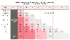 Figure 60-9
Method of determining the percentage of airway obstruction.
After easy passage of an endotracheal tube, a manometer is placed at the connection
of the elbow of the anesthesia circuit and the endotracheal tube. A stethoscope
is placed over the larynx and the circuit slowly pressurized. The pressure at which
a leak is auscultated is matched with the age of the patient and the size endotracheal
tube to assess the degree of laryngeal narrowing. It should be noted that this chart
is based on one institution's experience and the manufacturer of the endotracheal
tubes was not described; thus, the actual external diameter of the endotracheal tubes
used is unknown. (Redrawn with modification from Myer CM 3d, O'Connor DM,
Cotton RT: Proposed grading system for subglottic stenosis based on endotracheal
tube sizes. Ann Otol Rhinol Laryngol 103:319–323, 1994.)
Figure 60-9
Method of determining the percentage of airway obstruction.
After easy passage of an endotracheal tube, a manometer is placed at the connection
of the elbow of the anesthesia circuit and the endotracheal tube. A stethoscope
is placed over the larynx and the circuit slowly pressurized. The pressure at which
a leak is auscultated is matched with the age of the patient and the size endotracheal
tube to assess the degree of laryngeal narrowing. It should be noted that this chart
is based on one institution's experience and the manufacturer of the endotracheal
tubes was not described; thus, the actual external diameter of the endotracheal tubes
used is unknown. (Redrawn with modification from Myer CM 3d, O'Connor DM,
Cotton RT: Proposed grading system for subglottic stenosis based on endotracheal
tube sizes. Ann Otol Rhinol Laryngol 103:319–323, 1994.)
The approach to a difficult airway depends in part on whether it is a known difficult airway and previous records are available for review or the child has an unexpectedly difficult airway.[266] In the former situation, a difficult-airway cart with age- and size-appropriate airway equipment ( Table 60-7 ) is brought into the operating room, and the assistance of a colleague with pediatric airway skills is arranged.[267] In the latter situation, the difficult-airway cart may need to be brought into the room and skilled help summoned. The most common errors that I have observed in a number of malpractice cases are lack of equipment or inappropriate equipment, inadequate skilled help, and failure of the anesthesiologist to attempt cricothyrotomy soon enough. This is the reason that I emphasize the importance of having a dedicated pediatric difficult-airway cart, the need to be familiar with its contents and how to use the devices that it contains, and the availability of skilled help.
A child facing a potentially difficult laryngoscopic procedure should have, at most, light sedation before induction of anesthesia by mask to maintain spontaneous respirations. In this way the airway can be secured before "burning bridges" by administering a muscle relaxant and losing the help of spontaneous respirations. With the patient breathing spontaneously, the breath sounds can be used to guide successful tracheal intubation. When normal airway anatomy cannot be confirmed visually, placing a stylet in the endotracheal tube and bending the tip of the tube acutely (90-degree bend) allows placement of the tip of the tube in the midline just posterior to the epiglottis. One can then listen for breath sounds exiting from the end of the endotracheal tube, and once good air exchange is heard, the tube is advanced off the stylet (not inserted with the stylet). This technique allows advancement of the endotracheal tube at an acute angle without causing trauma to the anatomic structures. If the patient is not breathing spontaneously, this advantage may be lost. Spontaneous respirations with a facemask designed to allow fiberoptic intubation is another approach.[268] Having a skilled assistant pull the tongue forward out of the mouth with dry gauze or a plastic retractor will also aid visualization of the glottic opening. Fiberoptic intubation
| Equipment Category | Recommended Equipment |
|---|---|
| Airways | Oral and nasopharyngeal (trumpet) airways in all sizes for premature infants to adults |
| Endotracheal tubes | Cuffed and uncuffed endotracheal tubes of various sizes (uncuffed down to size 2.0 internal diameter [ID]) |
| Stylets | Stylets in several sizes |
| Laryngoscopy | Laryngoscope blades in multiple sizes and configurations |
|
|
Several handles, extra batteries |
|
|
Oxyscope * |
| Laryngeal mask airways | All sizes from 1.0 to 6.0 |
|
|
Large-volume syringes for the larger masks |
| Fiberoptic intubation | Fiberoptic scopes—several sizes, including one that will fit through a 2.5-mm-ID endotracheal tube |
|
|
Light source |
|
|
Teeth protectors |
|
|
Oral airways designed for fiberoptic intubation |
|
|
Silicon lubricant |
| Retrograde equipment | Retrograde intubation equipment (needle with guidewire) |
| Endotracheal tube exchangers | Various sizes for the full array of pediatric patients |
| Cricothyrotomy equipment | 1. Percutaneous cricothyrotomy kits designed for older and larger patients (needles, guidewire, and dilator) |
|
|
2. Jet ventilation-type devices, which are similar to an IV catheter (18, 14, and 13 gauge) and useful in infants |
| Jet ventilation equipment | Jet ventilation stylets and jet injector with appropriate hookup for wall oxygen |
| Carbon dioxide detector | For use in non-operating room venues |
If the patient should progress to a "can't ventilate/can't intubate"
situation, one must immediately establish a surgical airway. It is vital to properly
position the patient with a roll of towels under the shoulders to force the larynx
anteriorly so that the best access to the larynx is provided. It is also important
to have a variety of cricothyrotomy devices available because some are appropriate
for infants whereas others are appropriate only for older children and teenagers.
Devices appropriate for older patients include some using the Seldinger technique
(cricothyroid puncture, passing a guidewire, skin incision, then passing a dilator/catheter
over the wire [e.g., the Patil, Arndt, and Melker kits, Cook Critical Care, Inc.,
Bloomington, IN]) or the use of an armored transtracheal catheter (Cook Critical
Care). The simplest cricothyrotomy device that can be used on patients of all ages,
including infants, is an intravenous catheter, which can then be interfaced with
the 15-mm connector of a 3-mm-internal diameter endotracheal tube.[272]
Alternatively, a preformed
| Laryngeal Mask Size | Patient Weight |
|---|---|
| 1 | ≤5 kg |
| 1.5 | 5–10 kg |
| 2 | 10–20 kg |
| 2.5 | 20–30 kg |
| 3 | 30–50 kg |
| 4 | 50–70 kg |
| 5 | 70–100 kg |
| 6 | >100 kg |
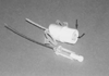 Figure 60-10
This device designed for transtracheal ventilation provides
one of the best systems for emergency airway management in a child with the "can't
ventilate/can't intubate" airway situation (Ventilation-Catheter, VBM Medical, Noblesville,
IN). Note that the design is similar to an intravenous catheter, which means that
the principles for use are familiar to all anesthesiologists and the device is small
enough to allow use even in neonates. In addition to a secure 15-mm adapter (straight
arrow) for interface with any standard bag ventilation device, a Luer-lok
connection (curved arrow) allows jet ventilation.
Figure 60-10
This device designed for transtracheal ventilation provides
one of the best systems for emergency airway management in a child with the "can't
ventilate/can't intubate" airway situation (Ventilation-Catheter, VBM Medical, Noblesville,
IN). Note that the design is similar to an intravenous catheter, which means that
the principles for use are familiar to all anesthesiologists and the device is small
enough to allow use even in neonates. In addition to a secure 15-mm adapter (straight
arrow) for interface with any standard bag ventilation device, a Luer-lok
connection (curved arrow) allows jet ventilation.
A child with intrathoracic airway obstruction has expiratory stridor and prolonged expiration (bronchiolitis, asthma, intrathoracic foreign body). In contrast, a child with extrathoracic upper airway obstruction has inspiratory stridor (epiglottitis, laryngotracheobronchitis, laryngeal foreign body). When agitated or crying, such patients have dynamic collapse of the airway ( Fig. 60-11 ), which can markedly worsen airway obstruction and lead to respiratory failure and hypoxemia. Therefore, events that can upset the child (drawing of blood for blood gas determination, venipuncture for blood tests, and separation from the parents) must be minimized.
I have found the following procedure effective for inducing anesthesia in a child with stridor. To minimize patient upset, the patient is brought to the operating room with the mother or father, who holds the child during the induction. Induction of anesthesia with halothane or sevoflurane in oxygen by mask is the preferred method because maintaining spontaneous respiration is critical. As soon as the child loses consciousness, the parent is escorted out of the operating room. With the patient lightly anesthetized and after infiltration of local anesthetic, an intravenous line is inserted to allow withdrawal of blood samples if indicated. Hydration is begun with lactated Ringer's solution (15 to 30 mL/kg), and atropine (0.02 mg/kg) is administered. At this point the plane of anesthesia can be deepened with greater safety. If stridor worsens or mild laryngospasm occurs, the pop-off valve is closed sufficiently to develop 10 to 15 cm H2 O of positive end-expiratory airway pressure. This procedure relieves most instances of airway obstruction caused by dynamic collapse of the airway and loss of pharyngeal muscle tone when the child attempts to inspire against an obstructed airway ( Fig. 60-12 ). As the level of anesthesia deepens, gentle assisting of ventilation may be necessary; however, maintaining spontaneous respiratory efforts if possible is important.
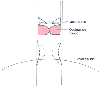 Figure 60-11
Infants and young children have highly compliant airway
structures. With normal respiration, some dynamic collapse of the extrathoracic
upper airway occurs (broken line). When a child
has upper airway obstruction (as in epiglottitis, laryngotracheobronchitis, and extrathoracic
foreign body) (shaded area) and struggles to breathe
against this obstruction, dynamic collapse of the trachea increases. This increase
in dynamic collapse (dotted line) augments mechanical
obstruction of the airway. Therefore, until the airway is secured, it is important
to avoid procedures that will upset the child. (Redrawn with modification
from Coté CJ, Todres ID: The pediatric airway. In
Coté CJ, Ryan JF, Todres ID, et al [eds]: A Practice of Anesthesia for Infants
and Children, 2nd ed. Philadelphia, WB Saunders, 1992, p 55.)
Figure 60-11
Infants and young children have highly compliant airway
structures. With normal respiration, some dynamic collapse of the extrathoracic
upper airway occurs (broken line). When a child
has upper airway obstruction (as in epiglottitis, laryngotracheobronchitis, and extrathoracic
foreign body) (shaded area) and struggles to breathe
against this obstruction, dynamic collapse of the trachea increases. This increase
in dynamic collapse (dotted line) augments mechanical
obstruction of the airway. Therefore, until the airway is secured, it is important
to avoid procedures that will upset the child. (Redrawn with modification
from Coté CJ, Todres ID: The pediatric airway. In
Coté CJ, Ryan JF, Todres ID, et al [eds]: A Practice of Anesthesia for Infants
and Children, 2nd ed. Philadelphia, WB Saunders, 1992, p 55.)
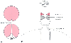 Figure 60-12
When a child has upper airway obstruction caused by laryngospasm
(A) or mechanical obstruction (B),
the application of approximately 10 cm H2
O of positive end-expiratory
pressure (PEEP) during spontaneous breathing often relieves the obstruction. That
is, PEEP helps keep the vocal cords apart (A) and
the airway open (B, broken lines).
If this simple maneuver does not relieve the obstruction, more vigorous positive-pressure
ventilation may be necessary. Airway obstruction caused by the tongue requires insertion
of an appropriately sized oral airway. (Redrawn with modification from Coté
CJ, Todres ID: The pediatric airway. In Coté
CJ, Ryan JF, Todres ID, et al [eds]: A Practice of Anesthesia for Infants and Children.
Philadelphia, WB Saunders, 1992, p 55.)
Figure 60-12
When a child has upper airway obstruction caused by laryngospasm
(A) or mechanical obstruction (B),
the application of approximately 10 cm H2
O of positive end-expiratory
pressure (PEEP) during spontaneous breathing often relieves the obstruction. That
is, PEEP helps keep the vocal cords apart (A) and
the airway open (B, broken lines).
If this simple maneuver does not relieve the obstruction, more vigorous positive-pressure
ventilation may be necessary. Airway obstruction caused by the tongue requires insertion
of an appropriately sized oral airway. (Redrawn with modification from Coté
CJ, Todres ID: The pediatric airway. In Coté
CJ, Ryan JF, Todres ID, et al [eds]: A Practice of Anesthesia for Infants and Children.
Philadelphia, WB Saunders, 1992, p 55.)
Any child with airway obstruction will have a long, slow induction of anesthesia before becoming sufficiently anesthetized to allow laryngoscopy and endotracheal intubation. Because a sevoflurane vaporizer only allows 2.5- to 3-MAC concentrations to be delivered whereas a halothane vaporizer allows 5- to 6-MAC concentrations, halothane would be my drug of choice in this circumstance. In addition, the slower excretion of halothane than that of sevoflurane would also provide better conditions for airway manipulation for a longer time. The issue of a full stomach is secondary to the airway problem; rapid induction of anesthesia is contraindicated in these patients.
A child with laryngotracheobronchitis or epiglottitis usually requires an uncuffed endotracheal tube that is 0.5 to 1.0 mm (internal diameter) smaller than normal; the use of a stylet facilitates its insertion.
A difficult-airway cart should also be present in the room. The surgical team should be mobilized and prepared to perform an emergency tracheotomy should total airway obstruction occur and mask ventilation or endotracheal intubation not be possible. A more detailed discussion of airway problems is presented in Chapter 42 .
|
|
|
|
|
|
|
|
|
|
|
|
|