Pneumocephalus
The issue of pneumocephalus arises most often in connection with
posterior fossa craniotomies performed with a head-up posture because these operations
entail the probability that air will be retained within the cranium.[89]
[90]
During these procedures, air may enter the
supratentorial space, much as air enters an inverted pop bottle. Depending on the
relationship of the brainstem and temporal lobes to the incisura, the pressure in
the air collection may or may not be able to equilibrate with atmospheric pressure.
This phenomenon has relevance to the use of N2
O because any N2
O
that enters a trapped gas space will augment the volume of that space. In those
(probably uncommon) intraoperative circumstances where there is in fact a completely
closed intracranial gas space, the use of N2
O may result in an effect
comparable to that of an expanding mass lesion. We do not view N2
O as
absolutely contraindicated because before dural closure, intracranial gas is probably
only rarely trapped. Nonetheless, attention to this possibility is important when
one is presented with the problem of an increasingly "tight" brain during a posterior
fossa craniotomy.[91]
[92]
During a posterior fossa procedure performed in a head-up posture,
when surgical closure has reached a stage such that the intracranial space has been
completely sealed from the atmosphere, it is probably appropriate to then omit N2
O
because of the possibility of contributing to tension pneumocephalus. Note that
the use of N2
O up to the point of dural closure may actually represent
a clinical advantage[93]
in that the gas pocket
can be expected to shrink more rapidly as a result of the presence of N2
O
(because N2
O will diffuse out much more quickly than nitrogen). Tension
pneumocephalus is often naively viewed as being exclusively a function of the use
of N2
O. However, it can most certainly occur as a complication of intracranial
neurosurgery entirely unrelated to the use of N2
O.
[94]
Tension pneumocephalus is one of the causes
of delayed awakening or nonawakening after both posterior fossa and supratentorial
procedures ( Fig. 53-9
).
[94]
[95]
It occurs
because air enters the cranium with the patient in a head-up position at a time when
the volume of the intracranial contents has been reduced as a result of some combination
of hypocapnia, good venous drainage, osmotic diuresis, and loss of CSF from the operative
field. When the cranium is closed and the patient is returned to the near-supine
position, CSF, venous blood volume, and extracellular fluid return or reaccumulate
and the air pocket becomes an unyielding mass lesion (because of the very slow diffusion
of nitrogen). It may cause delayed recovery of consciousness or severe headache.
Among supratentorial
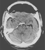 Figure 53-9
Postoperative computed tomographic scan demonstrating
a large pneumocephalus after a subfrontal approach to a suprasellar glioma. Immediately
postoperatively, the patient was confused and agitated and complained of severe headache.
Figure 53-9
Postoperative computed tomographic scan demonstrating
a large pneumocephalus after a subfrontal approach to a suprasellar glioma. Immediately
postoperatively, the patient was confused and agitated and complained of severe headache.
craniotomies, the largest residual air spaces occur after frontal skull base procedures
in which energetic brain relaxation measures are used to facilitate subfrontal access
(see Fig. 53-9
). At the
end of these procedures, which are typically performed in a supine/brow-up position,
it is not feasible to fill the intracranial dead space with normal saline as is commonly
done with smaller craniotomy defects, and a large residual pneumatocele may be left.
Once again, we doubt that the possible occurrence of this phenomenon represents
a contraindication to the use of N2
O. However, withdrawal of N2
O
may be appropriate at the time of cranial closure. The diagnosis of pneumocephalus
is confirmed by a brow-up lateral radiograph or CT scan. The treatment is a twist
drill hole followed by needle puncture of the dura.
Residual intracranial air should be considered at the time of
repeat anesthesia, neurosurgical or non-neurosurgical. Air frequently remains evident
on CT for more than 7 days after a craniotomy.[96]
Pneumocephalus can also develop de novo in the postoperative period in patients
who have a residual dural defect and communication between the nasal sinuses and
the intracranial space.[97]