|
|
|
|
|
|
|
|
|
|
|
|
|
|
|
Of the newer techniques available for monitoring patients during pediatric cardiovascular surgery, the most promising is echo-Doppler. Several reports have described the usefulness of intraoperative echo-Doppler during congenital heart surgery.[43] [44] [45] Two-dimensional echocardiography combined with pulsed-wave Doppler ultrasonography and color flow mapping is able to provide detailed morphologic as well as physiologic information in the majority of operative cases. Using echo-Doppler in the operating room, anatomic and physiologic data can be obtained prior to CPB, thus refining the operative plans. Pre-bypass echo-Doppler precisely defines anesthetic and surgical management.[16] [44] Because of the unrestricted epicardial and transesophageal echocardiography (TEE) approaches in anesthetized patients, new findings are frequently discovered and management plans changed accordingly ( Fig. 51-4 ).
Post-bypass echo-Doppler evaluation is able to immediately assess the quality of the surgical repair as well as assess cardiac function by examining ventricular wall motion and systolic thickening.[16] [44] This technique can show residual structural defects after bypass, which can be immediately repaired in the same operative setting and avoids leaving the operating room with significant residual structural defects that will require reoperation at a later time ( Fig. 51-5 ). By identifying patients with new RV and LV contraction abnormalities after bypass, as
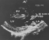 Figure 51-4
Intraoperative pre-cardiopulmonary bypass epicardial
echocardiogram in the long-axis view. Note the insertion of the papillary muscle
of the tricuspid valve on the interventricular septum. Based on this view, the surgeon
decided that ventricular septal defect (VSD) closure was possible in a child thought
preoperatively to be only a candidate for palliation. LA, left atrium; LV, left
ventricle; RV, right ventricle.
Figure 51-4
Intraoperative pre-cardiopulmonary bypass epicardial
echocardiogram in the long-axis view. Note the insertion of the papillary muscle
of the tricuspid valve on the interventricular septum. Based on this view, the surgeon
decided that ventricular septal defect (VSD) closure was possible in a child thought
preoperatively to be only a candidate for palliation. LA, left atrium; LV, left
ventricle; RV, right ventricle.
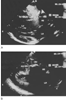 Figure 51-5
A, Echocardiogram with
a Doppler flow map in the long-axis view illustrating a residual ventricular septal
defect (VSD) resulting from patch dehiscence after initial repair. Turbulent flow
through the VSD appears as a mosaic of white particles (arrow).
This finding necessitated immediate reinstitution of cardiopulmonary bypass and
re-repair. B, Repeat Doppler flow map in the long-axis
view illustrates patch closure (arrow) of the VSD
after re-repair. Note the absence of turbulent flow with the loss of the mosaic
of white particles.; Ao, aorta; LA, left atrium; LV, left ventricle; RV, right ventricle.
Figure 51-5
A, Echocardiogram with
a Doppler flow map in the long-axis view illustrating a residual ventricular septal
defect (VSD) resulting from patch dehiscence after initial repair. Turbulent flow
through the VSD appears as a mosaic of white particles (arrow).
This finding necessitated immediate reinstitution of cardiopulmonary bypass and
re-repair. B, Repeat Doppler flow map in the long-axis
view illustrates patch closure (arrow) of the VSD
after re-repair. Note the absence of turbulent flow with the loss of the mosaic
of white particles.; Ao, aorta; LA, left atrium; LV, left ventricle; RV, right ventricle.
Two techniques for intraoperative echo-Doppler have been described: epicardial and TEE. Using TEE, the probe is placed after induction of anesthesia and intubation and is then available for monitoring of the patient. The advantage of this technique is its utility as a continuous monitor of cardiac structure and function, without interrupting surgery.[16] [47] Because of its ideal imaging location, TEE has been especially helpful in evaluating pulmonary venous return and the integrity of the left AV valve following mitral valvuloplasty, complete AV valve repair, and correction of complex congenital heart disease. Early limitation in views has been virtually eliminated as a result of clinical experience and improved biplane images. Pediatric biplane TEE probes have extended the patient weight limits to neonates between 2.5 and 3 kg.[48] Potential hazards of TEE that merit particular vigilance include descending aorta and airway compression due to probe size or during probe flexion. If a pre-repair TEE is indicated, the probe is removed during the procedure because of concerns about esophageal damage during hypothermia and low or no flow states.
A second technique for intraoperative echocardiographic analysis in children is the epicardial approach.[17] [44] [48] This approach requires passing a clean, short-focused 5.0- or 7.0-MHz transducer over the anesthesia screen into a sterile sheath, where it then can be placed on the epicardial surface of the heart. This technique best facilitates the probe manipulations necessary for thorough interrogation of the major structures and dynamic function of the heart. The advantage of this approach is that all views can be obtained in patients of any size. Among the disadvantages are the need for sufficient operator skill and experience to perform the manipulations, [46] the need to interrupt surgery to manipulate the probe, and the possible deleterious impact of direct myocardial mechanical manipulation. With the current TEE capabilities, epicardial imaging is rarely employed.
The primary goal of brain monitoring is to improve our understanding of cerebral function and dysfunction during cardiac surgery so that effective brain protection strategies can be developed. Because many of the determinants of normal brain perfusion become externally controlled by the cardiac team during CPB, such as flow rate (cardiac output), perfusion pressure, temperature, hematocrit, and PaCO2 , a knowledge of the effect of these factors on the brain in neonates, infants, and children is essential. Furthermore, the examination of the brain under unusual biologic circumstances, such as after deep hypothermic circulatory arrest (DHCA) or during continuous-flow CPB at deep hypothermia (18°C), permits a unique opportunity to describe cerebrovascular physiology and pathophysiology. Processed electroencephalography, transcranial Doppler imaging (TCD), cerebral blood flow (CBF), and metabolism measurements have provided important information during pediatric cardiovascular surgery.
Electroencephalography is helpful in monitoring physiologic functions of the central nervous system during deep hypothermic bypass and total circulatory arrest. For example, during deep hypothermia and before total circulatory arrest, the processed electroencephalogram can identify residual cerebral electrical activity. [49] Isoelectric silence can then be induced by further cooling, and any further brain activity detected by electroencephalography. Since this residual electrical activity during arrest is
TCD is one of a number of methods currently being used to monitor CBF during pediatric cardiac surgery.[50] [51] TCD technology uses the Doppler principle to detect shifts in the frequency of reflected signals from blood in the middle cerebral artery to calculate blood flow velocity. [52] Because the diameter of this large cerebral artery is relatively constant, flow velocity should approximate that of CBF. TCD has several advantages: (1) it is noninvasive, (2) it does not require radiation exposure, and (3) it is a continuous monitor. An additional advantage of this technique is the capability of assessing rapid alterations in blood flow velocity caused by temperature or perfusion changes, as commonly occur during cardiac surgery. The limitations of TCD monitoring include (1) reproducibility, especially at low flows, where minute movement of the patient's head can dramatically alter signal intensity and alter baseline measurements, and (2) the lack of validating studies of TCD during hypothermic CPB, when temperature, reduced flow rates, and the laminar flow characteristics of nonpulsatile perfusion may limit the accuracy of CBF velocity measurements. CBF velocity measurements by TCD have reasonable correlation with more standard measures of CBF during normothermia, and some studies have examined its validity during hypothermic CPB.[53]
TCD has been used to investigate the effect of CPB and deep hypothermic circulatory arrest on cerebral hemodynamics in children, as well as to assess the incidence of cerebral emboli. Recent studies examining the brain using TCD have enabled several investigative groups to provide important information regarding questions of normal and abnormal brain perfusion during cardiac surgery in children. Questions regarding cerebral perfusion pressure, autoregulation, effect of PaCO2 , and temperature have been addressed using TCD in children and are discussed subsequently. [50] [51] [53] This technique has also provided qualitative information regarding the presence of gaseous emboli in the middle cerebral artery during cardiac surgery.[54] [55] Quantification of this important mechanism of cerebral injury during cardiac surgery would be instructive. Future investigations using TCD should address this mechanism of injury as well.
CBF studies using xenon clearance technology have improved the understanding of cerebrovascular dynamics in young children during CPB and especially during deep hypothermia and after periods of circulatory arrest.[56] [57] [58] [59] [60] In general, this investigational tool has described the effects of CPB, temperature, and various perfusion techniques on CBF and, indirectly, on brain metabolism ( Fig. 51-6 ). Studies using this methodology have shown that some of the mechanisms of CBF autoregulation, such as pressure-flow
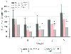 Figure 51-6
Bar chart of the changes in cerebral blood flow (CBF)
before, during, and after cardiopulmonary bypass (CPB) in 67 infants and children
(mean ± SD). Group A underwent repair with moderate hypothermic bypass (MoCPB)
at 28 to 32°C; group B, with deep hypothermic bypass (DHCPB) at 18 to 22°C;
and group C, with total circulatory arrest at 18°C. Note the impaired cerebral
reperfusion after total circulatory arrest (group C). Stage I, pre-bypass; stages
II and III, during hypothermic bypass; stage IV, rewarmed on bypass; stage V, after
bypass. (From Greeley WJ, Brusino FG, Ungerleider RM et al: The effects
of cardiopulmonary bypass on cerebral blood flow in neonates, infants, and children.
Circulation 80:1209, 1989.)
Figure 51-6
Bar chart of the changes in cerebral blood flow (CBF)
before, during, and after cardiopulmonary bypass (CPB) in 67 infants and children
(mean ± SD). Group A underwent repair with moderate hypothermic bypass (MoCPB)
at 28 to 32°C; group B, with deep hypothermic bypass (DHCPB) at 18 to 22°C;
and group C, with total circulatory arrest at 18°C. Note the impaired cerebral
reperfusion after total circulatory arrest (group C). Stage I, pre-bypass; stages
II and III, during hypothermic bypass; stage IV, rewarmed on bypass; stage V, after
bypass. (From Greeley WJ, Brusino FG, Ungerleider RM et al: The effects
of cardiopulmonary bypass on cerebral blood flow in neonates, infants, and children.
Circulation 80:1209, 1989.)
There now exists the capability of measuring cerebral metabolic activity during cardiac surgery. Methods for monitoring cerebral metabolic activity include determining the cerebral metabolic rate for oxygen (CMRO2 ), jugular venous bulb saturation, and near-infrared spectroscopy (NIRS). Using CMRO2 as a metabolic index, the effects of temperature and DHCA on brain metabolism have been described.[58] The primary effect of cooling during cardiac surgery is to reduce energy metabolism so that low-flow states and DHCA can be used.
Monitoring the efficacy of brain cooling can be performed by measuring the venous oxygen saturation of the brain. The higher the saturation level during cooling, the greater the oxygen metabolic suppression and the protective cooling effects. A catheter is easily placed in the right internal jugular vein and threaded retrograde to the venous bulb, positioned to assess the cerebral venous effluent. [60] Using a venous bulb catheter, potential mechanisms for brain injury have been identified and effective protection strategies developed. NIRS has the capability of measuring regional brain tissue oxyhemoglobin and cytochrome a,a3 , the terminal mitochondrial enzyme in the respiratory chain. Using NIRS, intracellular brain tissue oxygen delivery and utilization during CPB have been preliminarily observed.[62]
These growing improvements in our knowledge of the effects of CPB on the brain are of great importance because the last major area of morbidity associated with cardiac surgery is neurologic in origin.
|
|
|
|
|
|
|
|
|
|
|
|
|