Popliteal Fossa Block
The posterior muscles of the thigh are the biceps femoris, the
semimembranosus, semitendinosus, and the posterior portion of the adductor magnus.
As these muscles are traced distally from their origin on the ischial tuberosity,
they separate into medial (semimembranosus, semitendinosus) and lateral (biceps)
musculature, and they form the upper border of the popliteal fossa. The lower border
of the popliteal fossa is defined by the two heads of the gastrocnemius. In the
upper part of the popliteal fossa, the sciatic nerve lies posterolateral to the popliteal
vessels. The popliteal vein is medial to the nerve, and the popliteal artery is
most anterior, lying on the popliteal surface of the femur. Near the upper border
of the popliteal fossa, the two components of the sciatic nerve separate. The peroneal
nerve diverges laterally, and the larger tibial branch descends almost straight down
through the fossa. The tibial nerve and popliteal vessels then disappear deep to
the converging heads of the gastrocnemius muscle.
Clinical Applications
This block is chiefly used for foot and ankle surgery. The block
has also been successfully used in the pediatric population. Popliteal fossa block
is preferable to ankle block for surgical procedures requiring the use of a calf
tourniquet. The components of the sciatic nerve may be blocked at the level of the
popliteal fossa through posterior or lateral approaches. Supplemental block of the
saphenous nerve is required for surgical procedures to the medial aspect of the leg,
or when a calf tourniquet or Esmarch bandage is used.
Technique: Posterior Approach
The classic approach to the popliteal fossa is posteriorly, with
the patient positioned prone. However, access may also occur with the patient in
the lateral position (i.e., operative side nondependent) or supine position (i.e.,
with leg flexed at the hip and knee).
The borders of the popliteal fossa are identified by flexing the
knee joint. A triangle is constructed, with the base consisting of the skin crease
behind the knee, and the two sides composed of the semimembranosus (medially) and
the biceps (laterally). A bisecting line is drawn from the apex to the base of the
triangle, and a 5-cm needle is inserted at a site 5 to 10 cm above the skin fold
and 0.5 to 1 cm
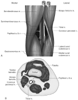 Figure 44-17
A, Anatomic landmarks
for the posterior approach to the sciatic nerve in the popliteal fossa (see Plate
14A
in the color atlas of this volume). B,
Anatomic landmarks for the lateral approach to the sciatic nerve in the popliteal
fossa (see Plate 14B
in
the color atlas of this volume).
Figure 44-17
A, Anatomic landmarks
for the posterior approach to the sciatic nerve in the popliteal fossa (see Plate
14A
in the color atlas of this volume). B,
Anatomic landmarks for the lateral approach to the sciatic nerve in the popliteal
fossa (see Plate 14B
in
the color atlas of this volume).
lateral to the bisecting line ( Fig.
44-17A
; see Plate 14A
in the color atlas of this volume). Classically, the 5-cm distance was described.
[48]
However, in an attempt to block the sciatic
nerve before its division, a 7- to 10-cm distance has been recommended.[49]
The needle is advanced at a 45-degree angle until a paresthesia or nerve stimulator
response is elicited. With a nerve stimulator technique, inversion is the motor
response that best predicts complete neural block of the foot.[50]
Injection of approximately 30 mL of local anesthetic solution is sufficient.
The success rate is typically 90% to 95%.[48]
[50]
No formal comparison between paresthesia and
nerve stimulator techniques has been performed to assess efficacy and complications.
It is believed that incomplete block is the result of poor diffusion (because of
the size of the sciatic nerve), the separate fascial coverings of the tibial and
peroneal nerves, or blockade of only a single component of the sciatic nerve. Identification
of the tibial and peroneal components decreases onset time and improves the success
rate.[51]
Technique: Lateral Approach
A lateral approach to blockade of the sciatic nerve in the popliteal
fossa has been described.[52]
Although block time
is somewhat longer, onset and quality of block are similar to the posterior approach.
[53]
The lateral approach allows the patient to
be positioned supine and eliminates the need for repositioning. The patient's leg
is extended, with the long axis of the foot at a 90-degree angle to the table. The
site of insertion is the intersection of the vertical line drawn from the upper edge
of the patella and the groove between the lateral border of the biceps femoris and
vastus lateralis. A 10-cm needle is advanced at a 30-degree angle posterior to the
horizontal plane (see Fig. 44-17B
;
see Plate 14B
in the color
atlas of this volume). Because the common peroneal nerve is located lateral to the
tibial nerve, the stimulating needle encounters the common peroneal nerve first with
the lateral approach. As with the classic posterior approach, an elicited inversion
response is sought.[50]
If a response associated
with common peroneal nerve stimulation (e.g., eversion) is elicited, the needle is
redirected more posteriorly.
Side Effects and Complications
As with other peripheral nerve blocks, neuropathy is the most
common complication. Intravascular injection may occur as a result of the presence
of vascular structures
within the popliteal fossa. Performance of popliteal fossa block in patients with
previous total-knee arthroplasty or vascular bypass (femoral-popliteal) should be
done with care. However, there have been no cases of graft disruption or joint infections
related to needle placement in these patients.