|
|
|
|
|
|
|
|
|
|
|
|
|
|
|
Four of the five individual nerves that can be blocked at the ankle to provide anesthesia of the foot are terminal branches of the sciatic nerve: the posterior tibial, sural, superficial peroneal, and deep peroneal branches. The sciatic nerve divides at or above the apex of the popliteal fossa to form the common peroneal and tibial nerves. The common peroneal nerve descends laterally around the head of the fibula, where it divides into the superficial and deep peroneal nerves.
The tibial nerve divides into the posterior tibial and sural nerves in the lower leg. The posterior tibial nerve becomes superficial at the medial border of the Achilles tendon near the artery of the same name, and the sural nerve emerges lateral to the Achilles tendon.
Ankle blocks are simple to perform and offer adequate anesthesia for surgical procedures of the foot not requiring a tourniquet above the ankle.
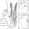
The posterior tibial nerve can be blocked with the patient in either the prone or the supine position. The posterior tibial artery is palpated, and a 25-gauge, 3-cm needle is inserted posterolateral to the artery at the level of the
 Figure 44-18
A, Anatomic landmarks
for a block of the posterior tibial and sural nerves at the ankle (see Plate
14A
in the color atlas of this volume). B,
Posterior tibial nerve and method of needle placement for a block at the ankle (see
Plate 14B
in the color
atlas of this volume). C, Sural nerve and method
of needle placement for a block at the ankle (see Plate
14C
in the color atlas of this volume).
The sural nerve is located superficially between the lateral malleolus and the Achilles tendon. A 25-gauge, 3-cm needle is inserted lateral to the tendon and is directed toward the malleolus as 5 to 10 mL of solution is injected subcutaneously (see Fig. 44-18A and C ; see Plate 15 in the color atlas of this volume). This block provides anesthesia of the lateral foot and the lateral aspects of the proximal sole of the foot.
The deep peroneal, superficial peroneal, and saphenous nerves can be blocked through a single needle entry site ( Fig. 44-19 ; see Plate 16 in the color atlas of this volume). A line is drawn across the dorsum of the foot connecting the malleoli. The extensor hallucis longus tendon is identified by having the patient dorsiflex the big toe. The anterior tibial artery lies between this structure and the tendon of the extensor digitorum longus muscle and is palpable at this level. A skin wheal is raised just lateral to the pulsation between the two tendons on the intermalleolar line. A 25-gauge, 3-cm needle is advanced perpendicular to
 Figure 44-19
A, Anatomic landmarks
for a block of the deep peroneal, superficial peroneal, and saphenous nerves at the
ankle (see Plate 16
in
the color atlas of this volume). B, Method of needle
placement for a block of the deep peroneal, superficial peroneal, and saphenous nerves
through a single needle entry site.
The needle is directed laterally through the same skin wheal while injecting 3 to 5 mL of solution subcutaneously, blocking the superficial peroneal nerve and resulting in anesthesia of the dorsum of the foot, excluding the first interdigital cleft. The same maneuver can be performed in the medial direction, thereby anesthetizing the saphenous nerve, a terminal branch of the femoral nerve that supplies a strip along the medial aspect of the foot.
Multiple injections are required for some techniques, resulting in discomfort for the patient. Persisting paresthesias may occur, but they are self-limited. The presence of edema or induration in the area of the ankle block can make palpation of landmarks difficult. Intravascular injection is possible but unlikely if aspiration for blood is negative. The volume of local anesthetic used is small, thereby decreasing the risk of local anesthetic toxicity.
|
|
|
|
|
|
|
|
|
|
|
|
|