|
|
|
|
|
|
|
|
|
|
|
|
|
|
|
The sciatic nerve (L4 and L5, S1 through S3) is the largest of the four peripheral nerves of the lower extremity, with a width of 2 cm as it leaves the pelvis with the posterior cutaneous nerve of the thigh. The sciatic nerve is composed of two nerves bound by a common sheath of connective tissue; the tibial component is medial and anterior, and the common peroneal component is lateral and slightly posterior. After passing through the sacrosciatic foramen beneath the piriformis muscle, it lies between the greater trochanter of the femur and the ischial tuberosity.
 Figure 44-14
Patient positioning for the posterior approach to the
sciatic nerve (see Plate 13A
in the color atlas of this volume).
Figure 44-14
Patient positioning for the posterior approach to the
sciatic nerve (see Plate 13A
in the color atlas of this volume).
Because of its wide sensory distribution, the sciatic nerve block can be used, together with a saphenous or femoral nerve block, for any surgical procedure below the knee that does not require a thigh tourniquet. It can also be combined with other peripheral nerve blocks to provide anesthesia for surgical procedures involving the thigh and knee. This form of anesthesia avoids the sympathectomy associated with neuraxial blocks and therefore may be advantageous when any shift in hemodynamics could be deleterious, such as in patients with significant aortic stenosis.
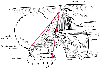 Figure 44-15
Anatomic landmarks for the posterior approach to a sciatic
nerve block (see Plate 13B
in the color atlas of this volume).
Figure 44-15
Anatomic landmarks for the posterior approach to a sciatic
nerve block (see Plate 13B
in the color atlas of this volume).
For the classic (posterior) approach of Labat, the patient is positioned laterally, with the leg to be blocked rolled forward onto the flexed knee as the heel rests on the knee of the dependent (nonoperative) leg ( Fig. 44-14 ; see Plate 13A in the color atlas of this volume).[6] A line is drawn to connect the posterior superior iliac spine to the greater trochanter of the femur. A perpendicular line is drawn bisecting this line and extending 5 cm caudad. A second line is drawn from the greater trochanter to the sacral hiatus. The intersection of this line with the perpendicular line indicates the point of needle entry and falls 3 to 5 cm along the line. A 22-gauge, 10- to 12-cm needle is advanced until a paresthesia or nerve stimulator response is elicited or bone is contacted ( Fig. 44-15 ; see Plate 13B in the color atlas of this volume). If bone is encountered, the needle is redirected systematically in a lateral or medial direction. After the needle is properly placed, a total of 20 to 30 mL of solution is injected.
The anterior approach is useful when the patient cannot be positioned for the classic posterior approach because of pain or lack of cooperation.[45] Initial blockade of the femoral nerve decreases the pain associated with this approach.
With the patient in the supine position, a line drawn along the inguinal ligament from the anterior superior iliac crest to the pubic tubercle is trisected. A second line parallel to the inguinal ligament is drawn, beginning at the tuberosity of the greater trochanter. A 22-gauge, 10.5-to 12-cm needle is inserted perpendicularly with a slightly lateral angulation at the point where the line representing the juncture of the middle and medial thirds crosses the second line. This needle is advanced until it contacts bone, the lesser trochanter of the femur ( Fig. 44-16 ). The needle is redirected medially past the femur, and a paresthesia or nerve stimulator response is sought at a depth of about 5 cm past the bone. A total of 20 to 25 mL of solution is injected incrementally after careful aspiration.
The sciatic nerve can also be blocked with the patient in the lateral[46] and lithotomy positions,[47] although these are rarely employed clinically.
Many methods have been tried to improve success with sciatic nerve blockade. Attempts to place the needle in
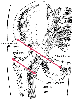 Figure 44-16
Anatomic landmarks for the anterior approach to a sciatic
nerve block.
Figure 44-16
Anatomic landmarks for the anterior approach to a sciatic
nerve block.
The block is technically difficult to perform and can be quite painful. Hematoma formation is possible. The risk of nerve damage is also reported, although persistent paresthesias are usually self-limited. A minimal degree of vasodilation may occur with sciatic nerve block.
|
|
|
|
|
|
|
|
|
|
|
|
|