Caudal Technique
Caudal anesthesia requires identification of the sacral hiatus.
The sacrococcygeal ligament (i.e., extension of ligamentum flavum) overlying the
sacral hiatus lies between the sacral cornu. To facilitate locating the cornu, the
posterior superior iliac spines should be located and, by using the line between
them as one side of an equilateral triangle, the location of the sacral hiatus approximated
( Fig. 43-18
). After the
sacral hiatus is identified, the index and middle finger of the palpating hand are
placed on the sacral cornu, and the caudal needle is inserted at an angle of approximately
45 degrees to the sacrum. While advancing the needle, a decrease in resistance to
needle insertion should be appreciated as the needle enters the caudal canal. The
needle is advanced until bone (i.e., dorsal aspect of the ventral plate of the sacrum)
is contacted and then slightly withdrawn, and the needle is redirected so that the
angle of insertion relative to the skin surface is decreased. In male patients,
this angle is almost parallel to the coronal plane; in female patients, a slightly
steeper angle (15 degrees) is necessary. During redirection of the needle and after
loss of resistance is again encountered, the needle is advanced approximately 1 to
2 cm into the caudal canal. Further advance is not attempted because dural puncture
and unintentional
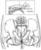 Figure 43-18
Caudal technique. A,
Palpating fingers locate the sacral cornua by using equilateral triangle (shown in
Fig. 43-5
). B,
Needle insertion is carried out by insertion and withdrawal in stepwise fashion (so-called
1-2-3-insertion), until needle can be advanced into the caudal canal and solution
can be injected easily (without creation of a subcutaneous "lump" of fluid).
Figure 43-18
Caudal technique. A,
Palpating fingers locate the sacral cornua by using equilateral triangle (shown in
Fig. 43-5
). B,
Needle insertion is carried out by insertion and withdrawal in stepwise fashion (so-called
1-2-3-insertion), until needle can be advanced into the caudal canal and solution
can be injected easily (without creation of a subcutaneous "lump" of fluid).
intravascular cannulation become more likely. One method of increasing the likelihood
of correct caudal needle placement is to inject 5 mL of saline rapidly through the
caudal needle while palpating the skin overlying the sacrum. If no midline bulge
is detected, the needle is probably correctly positioned. In contrast, if a midline
bulge is detected during saline injection, the needle is incorrectly positioned.
TABLE 43-8 -- Comparative onset times and analgesic durations of local anesthetics administered
epidurally in 20- to 30-mL volumes
|
|
|
Duration (min) |
|
Drug |
Conc. (%) |
Onset (min) |
Plain |
1:200,000 Epinephrine |
|
2-Chloroprocaine |
3 |
10–15 |
45–60 |
60–90 |
|
Lidocaine |
2 |
15 |
80–120 |
120–180 |
|
Mepivacaine |
2 |
15 |
90–140 |
140–200 |
|
Bupivacaine |
0.5–0.75 |
20 |
165–225 |
180–240 |
|
Etidocaine |
1 |
15 |
120–200 |
150–225 |
|
Ropivacaine |
0.75–1.0 |
15–20 |
140–180 |
150–200 |
|
Levobupivacaine |
0.5–0.75 |
15–20 |
150–225 |
150–240 |
|
Data from Cousins MJ, Bromage PR: Epidural neural
blockade. In Cousins MJ, Bridenbaugh PO (eds):
Neural Blockade in Clinical Anesthesia and Management of Pain. Philadelphia, JB
Lippincott, 1988, p 255 and other sources. |
After ensuring correct needle position and before injection of
the therapeutic dose of caudal anesthetic, aspiration and a test dose should be administered
because, as in lumbar epidural anesthesia, a vein or the subarachnoid space can be
entered unintentionally.