|
|
|
|
|
|
|
|
|
|
|
|
|
|
|
The technique of epidural anesthesia relies on the same four Ps as spinal anesthesia, with minor modifications.
In addition to patient preparation, selection of epidural equipment is essential. The physician must decide on a continuous or single-shot technique. This is the principal determinant of needle selection. If a single-shot epidural technique is chosen, a Crawford needle is appropriate; if a continuous catheter technique is indicated, a Tuohy or another needle with a laterally facing opening is chosen ( Fig. 43-15A and Fig. 43-15B ). A method of identifying the epidural space
The patient positions necessary for epidural puncture are the same as those for spinal anesthesia, with the exception of the prone position for the caudal approach. Inadequate positioning of the patient can negate otherwise meticulous technique and should be prevented. Limitation of block spread (i.e., height) is not clinically predictable with position alterations during epidural anesthesia because gravity and solution baricity are not intimately related to block spread. Despite these facts, Seow and coworkers[132] have demonstrated slightly faster block onset times in patients' dependent body regions when the lateral decubitus position was used for epidural block.
Three positions are available for caudal anesthesia, with the prone position most often chosen in adults, the lateral decubitus position in children, and the knee-chest position infrequently used. The lateral decubitus position is used in children because it is easier to maintain a patent airway in this position than in the prone position, and the landmarks are more easily palpable than in adults (see Chapter 45 ). This consideration is valuable because caudal anesthesia is often combined with general anesthesia in pediatric patients to decrease the amount of
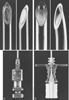 Figure 43-15a
Epidural needles with catheter assortment. A,
A 19-gauge, reusable Crawford epidural needle. B,
A 19-gauge, disposable Tuohy needle.
Figure 43-15a
Epidural needles with catheter assortment. A,
A 19-gauge, reusable Crawford epidural needle. B,
A 19-gauge, disposable Tuohy needle.
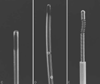 Figure 43-15b
C, Single-end-hole epidural
catheter. D, Closed- tip, multiple-side-hole catheter.
E, Spring-wire—reinforced, polymer-coated epidural
catheter.
Figure 43-15b
C, Single-end-hole epidural
catheter. D, Closed- tip, multiple-side-hole catheter.
E, Spring-wire—reinforced, polymer-coated epidural
catheter.
Epidural technique requires placement of the needle tip into the ligamentum flavum for loss-of-resistance and hanging-drop methods. Placing the needle (with stylet) into the ligamentum flavum before attaching the syringe or placing solution into the needle hub allows an improved appreciation of epidural anatomy for the operator. If the needle is merely inserted into the supraspinous ligament and then loss-of-resistance or hanging-drop insertion is begun, an increased chance of false release seems likely.
When a lumbar approach is used, the depth from skin to ligamentum flavum commonly approaches 4 cm, with most (80%) patients between 3.5 and 6 cm. In this region, the ligamentum flavum is 5 to 6 mm thick in the midline, requiring needle control if unintentional dural puncture is to be prevented. There are some exploring the use of ultrasound to help minimize dural puncture by more accurately defining depth of skin-to-epidural-space distance.[133] When a thoracic approach is chosen, control is of equal or greater importance because injury to the spinal cord is possible if the needle is advanced too far. Theoretically, the increased angle of needle insertion in the thoracic region may provide an element of safety because the more acute angle necessary to gain epidural cannulation provides some margin of safety ( Fig. 43-17 ). Clinically, thoracic epidural anesthetics do not appear to
 Figure 43-16
Prone position for caudal technique. A pillow is used
under the anterior iliac crests to rotate the pelvis, the legs are spread 20 degrees
to ease identification of the sacral hiatus, and the heels are rotated laterally
to relax the gluteal musculature.
Figure 43-16
Prone position for caudal technique. A pillow is used
under the anterior iliac crests to rotate the pelvis, the legs are spread 20 degrees
to ease identification of the sacral hiatus, and the heels are rotated laterally
to relax the gluteal musculature.
 Figure 43-17
A, Lumbar and thoracic
epidural technique. The increased angle of needle insertion during thoracic epidural
cannulation may provide a slightly longer distance of "needle travel" before entering
the subarachnoid space. B, in contrast to lumbar
epidural cannulation C, where the distance traveled
is modified by more perpendicular angle of needle insertion.
Figure 43-17
A, Lumbar and thoracic
epidural technique. The increased angle of needle insertion during thoracic epidural
cannulation may provide a slightly longer distance of "needle travel" before entering
the subarachnoid space. B, in contrast to lumbar
epidural cannulation C, where the distance traveled
is modified by more perpendicular angle of needle insertion.
The preferred method of carrying out the loss-of-resistance technique involves inserting the needle to the ligamentum flavum and then attaching a 3- to 5-mL glass syringe filled with 2 mL of saline and a small (0.25-mL) air bubble. The needle is grasped with the nondominant hand and pulled toward the epidural space, while the dominant hand (thumb) applies constant steady pressure on the syringe plunger, compressing the air bubble. When the epidural space is entered, the pressure applied to the syringe plunger allows the solution to flow without resistance into the epidural space (see Plate 7A in the color atlas of this volume).
An alternative, although with a less precise end point, is the technique of hanging-drop identification of entry into the epidural space. After the needle is placed into the ligamentum flavum, a drop of solution is placed within the hub of the needle. When the needle is advanced into the epidural space, the solution should be "sucked in" (see Plate 7B in the color atlas of this volume). The theory behind this maneuver has been attributed to a subatmospheric pressure in the epidural space. The subatmospheric pressure has been related to the expansion of the epidural space as the needle pushes the dura away from the ligamentum flavum.[135] The negative intrathoracic pressure may influence the pressure in the epidural space in the thoracic region.
Regardless of the method selected for needle insertion, when the anesthesiologist chooses to cannulate the epidural space with a catheter, success may be increased by advancing the needle 1 to 2 mm after the space is identified. The incidence of unintentional intravenous cannulation with an epidural catheter may be lessened by injecting air or solution before threading the catheter.[136] [137] Unless radiographic guidance is used for some special reason, epidural catheters should be inserted only 2 to 3 cm into the epidural space for surgical patients or obstetric patients needing rapid onset of analgesia. [138] Threading more catheter may increase the likelihood of catheter malposition. If malposition does occur, data suggest that one of the more common sites of malposition is entry of the catheter into the anterior epidural space.[139] In obstetric patients, it appears that catheters should be inserted between 4 and 6 cm to optimize efficacy and prevent unintentional movement of the catheters during prolonged labor analgesia.[140] [141] Multiport (three lateral ports) or single-end-hole (uniport or distal port) catheters are available. The choice is practically up to individual clinicians, although it has been shown in laboring patients that multiport catheters reduce the incidence of inadequate analgesia compared with uniport catheters. [142]
Despite an adequately positioned catheter during first use of local anesthetic, each subsequent injection should be preceded by aspiration and an epidural test dose because catheter migration into vessels and subarachnoid or subdural spaces does occur. In the past, many considered the catheter unusable in the event that the epidural catheter hub becomes disconnected from the epidural catheter. It appears that the catheter can be reused if strict reconnection criteria are followed. [143]
|
|
|
|
|
|
|
|
|
|
|
|
|