SITES OF NERVE STIMULATION AND DIFFERENT MUSCLE RESPONSES
In principle, any superficially located peripheral motor nerve
may be stimulated. In clinical anesthesia, the ulnar nerve is the most popular site;
the median, the posterior tibial, common peroneal, and facial nerves are also sometimes
used. For stimulation of the ulnar nerve, the electrodes are best applied at the
volar side of the wrist ( Fig. 39-9
).
The distal electrode should be placed about 1 cm proximal to the point at which
the proximal flexion crease of the wrist crosses the radial side of the tendon to
the flexor carpi ulnaris muscle. The proximal electrode preferably should be placed
2 to 5 cm proximal to the
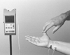 Figure 39-9
Evaluation of neuromuscular blockade by feeling the response
of the thumb to stimulation of the ulnar nerve. (Courtesy of Organon Ltd.,
Dublin, Ireland.)
Figure 39-9
Evaluation of neuromuscular blockade by feeling the response
of the thumb to stimulation of the ulnar nerve. (Courtesy of Organon Ltd.,
Dublin, Ireland.)
distal electrode. With this placement of the electrodes, electrical stimulation
normally elicits only finger flexion and thumb adduction. If the one electrode is
placed over the ulnar groove at the elbow, thumb adduction is often pronounced because
of stimulation of the flexor carpi ulnaris muscle. When this latter placement of
electrodes (sometimes preferred in small children) is used, the active negative electrode
should be at the wrist to ensure a maximal response. Polarity of the electrodes
is less crucial when both electrodes are close to each other at the volar side of
the wrist; however, placement of the negative electrode distally normally elicits
the greatest neuromuscular response.[34]
When the
temporal branch of the facial nerve is stimulated, the negative electrode should
be placed over the nerve, and the positive electrode should be placed somewhere else
over the forehead.
Because different muscle groups have different sensitivities to
neuromuscular blocking agents, results obtained for one muscle cannot be extrapolated
automatically to other muscles. The diaphragm is among the most resistant of all
muscles to both depolarizing[35]
and nondepolarizing
neuromuscular blocking drugs.[36]
In general, the
diaphragm requires 1.4 to 2.0 times as much muscle relaxant as the adductor pollicis
muscle for an identicald egree of blockade ( Fig.
39-10
).[36]
Also of clinical significance
are the facts that onset time is normally shorter for the diaphragm than for the
adductor pollicis muscle and that the diaphragm recovers from paralysis more quickly
than do the peripheral muscles ( Fig.
39-11
).[37]
The other respiratory
 Figure 39-10
Mean cumulative dose-response curve for pancuronium in
two muscles shows that the diaphragm requires approximately twice as much pancuronium
as does the adductor pollicis muscle for the same amount of neuromuscular blockade.
The depression in muscle response to the first stimulus in TOF nerve stimulation
(probit scale) was plotted against dose (log scale). Force of contraction of the
adductor pollicis was measured on a force-displacement transducer; response of the
diaphragm was measured electromyographically. (From Donati F, Antzaka C,
Bevan DR: Potency of pancuronium at the diaphragm and the adductor pollicis muscle
in humans. Anesthesiology 65:1, 1986.)
Figure 39-10
Mean cumulative dose-response curve for pancuronium in
two muscles shows that the diaphragm requires approximately twice as much pancuronium
as does the adductor pollicis muscle for the same amount of neuromuscular blockade.
The depression in muscle response to the first stimulus in TOF nerve stimulation
(probit scale) was plotted against dose (log scale). Force of contraction of the
adductor pollicis was measured on a force-displacement transducer; response of the
diaphragm was measured electromyographically. (From Donati F, Antzaka C,
Bevan DR: Potency of pancuronium at the diaphragm and the adductor pollicis muscle
in humans. Anesthesiology 65:1, 1986.)
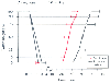 Figure 39-11
Evolution of twitch height (mean ± SD) of the
diaphragm (closed circles) and of the adductor pollicis
muscle (open circles) in 10 anesthetized patients
after administration of atracurium 0.6 mg/kg. (From Pansard J-L, Chauvin
M, Lebrault C, et al: Effect of an intubating dose of succinylcholine and atracurium
on the diaphragm and the adductor pollicis muscle in humans. Anesthesiology 67:326,
1987.)
Figure 39-11
Evolution of twitch height (mean ± SD) of the
diaphragm (closed circles) and of the adductor pollicis
muscle (open circles) in 10 anesthetized patients
after administration of atracurium 0.6 mg/kg. (From Pansard J-L, Chauvin
M, Lebrault C, et al: Effect of an intubating dose of succinylcholine and atracurium
on the diaphragm and the adductor pollicis muscle in humans. Anesthesiology 67:326,
1987.)
muscles are less resistant than the diaphragm, as are the larynx and the corrugater
supercilii muscles.[38]
[39]
[40]
[41]
[42]
[43]
Most sensitive are the abdominal muscles,
the
orbicularis oculi muscle, the peripheral muscles of the limbs, and the geniohyoid,
masseter, and upper airway muscles.[44]
[45]
[46]
[47]
[48]
From a practical clinical point of view, it is worth noting that (1) the corrugator
supercilii response to facial nerve stimulation reflects the extent of neuromuscular
blockade of the laryngeal adductor muscles (and the diaphragm?) better than does
the response of the adductor pollicis to ulnar nerve stimulation[38]
[39]
and (2) the upper airway muscles seem to be
more sensitive than peripheral muscles.[45]
[46]
Although three investigations using acceleromyography have indicated small differences
in the response to TOF nerve stimulation in the arm (adductor pollicis muscle) and
the leg (flexor hallucis brevis muscle), these differences are probably of little
clinical significance.[49]
[50]
[51]
[52]
The precise source of these differences is unknown. Possible
causes may be differences in acetylcholine receptor density, acetylcholine release,
acetylcholinesterase activity, fiber composition, innervation ratio (number of neuromuscular
junctions), blood flow, and muscle temperature.
In assessing neuromuscular function, the use of a relatively sensitive
muscle such as the adductor pollicis of the hand has both disadvantages and advantages.
Obviously, during surgery it is a disadvantage that even total elimination of the
response to single-twitch and TOF stimulation does not exclude the possibility of
movement of the diaphragm, such as hiccupping and coughing. PTC stimulation, however,
allows for evaluation of the very intense blockade necessary to ensure total paralysis
of the diaphragm. On the positive side, the risk of overdosing the patient decreases
if the response of a relatively sensitive muscle is used as a guide to the administration
of muscle relaxants during surgery. Also, during recovery, when the adductor pollicis
has recovered sufficiently, it can be assumed that no residual neuromuscular blockade
exists in the diaphragm or in other resistant muscles.