PREOPERATIVE EVALUATION OF RENAL FUNCTION
The greater the magnitude and duration of the surgical insult
and the number of acute and chronic risk factors, the greater is the likelihood of
perioperative renal compromise and the need for preoperative identification of renal
function (see Chapter 25
and Chapter 27
). In patients
with inadequate blood flow, injury is commonly caused by the added risk of drugs,
by abnormal hemodynamics, or by preexisting disease.[20]
Studies have shown that 12% to 14% of patients who develop acute renal insufficiency
disease during hospitalization do so after radiographic procedures with contrast.
If preexisting renal insufficiency exists, progressive deterioration in renal function
occurs 42% of cases, with a poor prognosis if dialysis is required.[78]
Acute risk factors, such as volume depletion, aminoglycoside use, radiocontrast
dye exposure, use of nonsteroidal anti-inflammatory drugs, septic shock, and pigmenturia,
augment the risk of ARF rather than induce it. It appears that the combined interaction
of these multiple acute risk factors is central in the pathogenesis of ARF ( Table
37-2
).[15]
Patients with preexisting
renal insufficiency, for example, are especially prone to develop ARF as a result
of cardiovascular surgery. Patients with diabetes mellitus and renal insufficiency
are especially vulnerable to radiocontrast agents. A review of the literature suggests
that the most critical determinants of postoperative renal function are preoperative
renal function, maintenance of appropriate intravascular volume, and normal myocardial
function.[13]
Although variables have been identified
that increase the likelihood of renal dysfunction outcomes, there remains unexplained
variability in prediction accuracy. Genetic polymorphism may predispose patients
to acute perioperative renal dysfunction, similar to the situation that has been
identified in chronic
renal disease. The apolipoprotein E4 was reported to be more associated with reduced
postoperative increases in serum creatinine after cardiac surgery compared with the
apolipoprotein E3 or E2 polymorphisms.[79]
Despite substantially reduced renal function, impaired functional
reserve capacity may only become evident when stress imposes severe demands on renal
function; therefore, a comprehensive evaluation of renal function reserve should
be sought before surgery. In addition to intrinsic renal disease, several extrinsic
variables influence the outcome of renal function tests: intracellular and extracellular
volume, cardiovascular function, and neuroendocrine factors.[80]
[81]
Advanced age also markedly decreases renal
function reserve. GFR, normally about 125 mL/min in a young adult, decreases to
about 80 mL/min at 60 years of age and to 60 mL/min at 80 years. Although not necessarily
the primary cause for end-stage renal disease, hypertension in up to 85% of patients
with renal failure is a major risk factor for the high cardiovascular morbidity that
occurs.[82]
In addition to its contribution to renal failure in hypertensive
nephrosclerosis, resulting from essential hypertension and hyperfiltration in diabetic
nephropathy, hypertension also contributes to the loss of renal function associated
with aging. Preoperative isolated systolic blood pressure (SBP) hypertension and
wide pulse pressure (i.e., the difference between SBP and diastolic blood pressure
[DBP]) are strong independent predictors of postoperative renal dysfunction.[10]
[11]
It appears that an increase in conduit vessel
stiffness with inadequate flow during low-pressure states contributes to increased
preoperative renal dysfunction and hemodialysis-dependent renal failure. Four groups
of preoperative hypertensive patients were identified in an analysis of 5065 patients
undergoing coronary artery bypass surgery. Isolated SBP (SBP > 160 mm Hg, DBP
< 90 mm Hg), isolated DBP (SBP < 160 mm Hg, DBP > 90 mm Hg), combined SBP
and DBP, and wide pulse pressure (> 80 mm Hg). In that analysis, the rate of
wide pulse pressure was 8%, and that of isolated SBP was 2.3%. Mild hypertensive
disease that is associated with renal disease therefore implies that renal disease
may be primary.
Reliable evaluation requires knowledge of various renal function
tests and their diagnostic limitations. For appropriate management, ischemic causes
(e.g., prerenal azotemia) should ideally be distinguished from factors that maintain
the intrinsic state of renal failure (e.g., acute tubular necrosis). Prerenal azotemia
and acute tubular necrosis, however, may not be mutually exclusive. Unfortunately,
there is no simple, inexpensive test that adequately quantifies renal function.
The readily available tests fail to accurately reflect the status of the kidneys
in a large percentage of patients, especially those who are elderly, malnourished,
or dehydrated. Commonly obtained tests of renal function are urinalysis, BUN determination,
serum creatinine determination, and creatinine clearance.
Urinalysis provides qualitative information that must be interpreted
cautiously. Hematuria (i.e., more than one or two red blood cells per high-power
field in a concentrated sediment) suggests glomerular disease or, in a trauma patient,
injury to the kidneys or the lower urinary tract. A urine test result that is positive
for blood in the absence of red blood cells suggests the presence of free hemoglobin
or myoglobin in the urine. Pyuria (i.e., more than four white blood cells per high-power
field) suggests urinary tract infection. Although urine may normally contain hyaline
and granular casts, cellular casts represent a pathologic condition. Red blood cell
casts suggest interstitial nephritis, including pyelonephritis. Urinary pH, although
difficult to interpret on a spot urine sample, may assist in the diagnosis of some
acid-base disturbances. The pH of urine tends to be more acidic when factors that
initiate failure are prerenal rather than postrenal. The presence of proteinuria
on a routine dipstick examination may be normal, or it may suggest severe renal disease.
In a concentrated urine sample, trace or 1+ proteinuria is a nonspecific finding,
whereas 3+ or 4+ proteinuria suggests glomerular disease. The existence of glucosuria
without hyperglycemia indicates proximal tubular damage. Lysozymuria, an increase
of the enzymatic protein lysozyme in urine, occurs if the serum level is elevated
above the normal renal threshold (45 mg/mL) or when renal tubular function is impaired.
[83]
The normal urinary lysozyme level is less
than
1.9 mg/mL, and a level greater than 5 mg/mL is regarded as evidence for significant
renal tubular damage. Elevated serum levels of lysozyme may be a consequence of
renal failure as well as a marker for it. Because white blood cells have a high
concentration of lysozyme, urinary tract infections may also lead to elevated serum
levels.
The BUN and serum creatinine levels offer rapid but inexact estimates
of creatinine clearance. Acute elevations of BUN and serum creatinine levels occur
in approximately 5% of all hospital admissions and in up to 20% of intensive care
patients.[16]
The incidence increases directly
with the severity of trauma and the degree of injury or disease. The incidence and
severity of ARF usually is greater when preoperative serum creatinine level is greater
than 2 mg/dL4
; however, an isolated serum creatinine measurement is an
unreliable indicator of GFR when renal function is changing. There is an inverse
logarithmic relationship between GFR and serum creatinine concentration ( Fig.
37-7
). Decreasing the rate by one half results in twice the serum creatinine
level. For example,
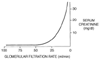 Figure 37-7
The inverse logarithmic relationship between serum creatinine
concentration (y axis) and glomerular filtration
rate (x axis). Notice that the serum creatinine
level is not markedly increased until there is a reduction in glomerular filtration
rate to about one fourth of normal (120 to 30 mL/min).
Figure 37-7
The inverse logarithmic relationship between serum creatinine
concentration (y axis) and glomerular filtration
rate (x axis). Notice that the serum creatinine
level is not markedly increased until there is a reduction in glomerular filtration
rate to about one fourth of normal (120 to 30 mL/min).
a patient with a preoperative serum creatinine level of 0.6 mg/dL whose filtration
rate was reduced by one half (e.g., after a large preoperative contrast dye load)
would have a serum creatinine level of 1.2 mg/dL, still in the normal range. A single
serum creatinine measurement is not a very sensitive test for preoperative renal
function reserve. However, it is relatively specific and useful in considering significant
renal disease; it would be rare for someone who is not emaciated to have a creatinine
level of 1.5 mg/dL and to have a creatinine clearance rate of less than 40 mL/min.
The measurement of creatinine clearance may constitute the best overall indicator
of GFR, although this index also has limitations. In one study, more than one half
of 131 critically ill patients had normal values for urine output, BUN, and serum
creatinine but had reduced creatinine clearnace. Creatinine clearance was the best
predictor of mortality among these patients compared with the other measures of renal
function.[84]
Although the serum creatinine level
may be a more reliable measure of glomerular function than the BUN level, the simultaneous
determination of BUN and serum creatinine levels offers a more complete evaluation
of renal function than either determination alone. Ordinarily, the BUN-to-creatinine
ratio is approximately 10, and if the BUN level is approximately 10 times as great
as the serum creatinine level, the clinician may conclude that the measurements are
probably correct. Conversely, if the ratio deviates significantly from 10, the clinician
should consider the nonrenal factors influencing the BUN level or serum creatinine
level, or both. All three measurements (i.e., BUN, serum creatinine level, and creatinine
clearance) require careful interpretation.