WAVEFORM ANALYSIS OF EXPIRED RESPIRATORY GASES
Capnography, the measurement of CO2
in expired gases,
has evolved in the last few years into a commonly used procedure. Whereas a variety
of techniques can be used for CO2
measurement (e.g., mass spectrometry,
Raman analysis), most capnographs rely on infrared absorption.[169]
Use of this technique can reliably and quantitatively provide vital respiratory
monitoring information in the operating room and in all critical care areas.
End-tidal PO2
(PETO2
)
may be used as an estimate of alveolar PO2
and therefore PaO2
. Whereas end-tidal
CO2
(PETCO2
) analysis has achieved
a high degree of popularity, this has not occurred for PO2
monitoring because of the variable A-a gradient.
In normal individuals, this gradient may be less than 10 mm Hg, but in patients
with severe V̇A/ mismatching, the gradient
may be substantially increased. The A-a gradient
is increased at high inspired O2
concentrations, even with normal lungs.
PETO2
therefore almost always overestimates
PaO2
. For example, the PETO2
of a cadaver being ventilated with 100% O2
would be approximately 700
mm Hg (Pbarometric
- PH2
O)!
Nevertheless, exhaled O2
analysis can be useful in monitoring the adequacy
of nitrogen washout in preparation for induction of general anesthesia, particularly
when a period of apnea is expected. Clinicians often select the time of induction
of anesthesia at a point when the inspired-expired percentage of O2
has
decreased to a plateau (typically less than 10%). An example of a continuous tracing
of expired PO2
, demonstrating nitrogen
washout, is shown in Figure 36-14
.
mismatching, the gradient
may be substantially increased. The A-a gradient
is increased at high inspired O2
concentrations, even with normal lungs.
PETO2
therefore almost always overestimates
PaO2
. For example, the PETO2
of a cadaver being ventilated with 100% O2
would be approximately 700
mm Hg (Pbarometric
- PH2
O)!
Nevertheless, exhaled O2
analysis can be useful in monitoring the adequacy
of nitrogen washout in preparation for induction of general anesthesia, particularly
when a period of apnea is expected. Clinicians often select the time of induction
of anesthesia at a point when the inspired-expired percentage of O2
has
decreased to a plateau (typically less than 10%). An example of a continuous tracing
of expired PO2
, demonstrating nitrogen
washout, is shown in Figure 36-14
.
According to the gas sampling technique, infrared CO2
monitors are in one of two categories: sidestream monitors, which draw a continuous
sample of the gas from the respiratory circuit into the measuring cell, and mainstream
monitors, which directly straddle the airway with a reading cell placed at the attachment
between respiratory circuit and endotracheal tube or breathing mask. The key difference
in use between the two types of capnographs depends on details of practical importance
and on the type and duration of the monitoring environment.
Sidestream Capnographs
Sidestream capnographs depend crucially on a sampling flow that
continuously aspirates from the side of the main respiratory gas flow a fixed amount
of gas. The rate of gas sampling can usually be adjusted from 50 to 500 mL/min and
sometimes up to 2 L/min. This continuous bias flow can be the source of significant
methodologic error. If the sampling flow ever exceeds the expired gas flow,
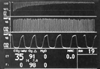 Figure 36-14
Monitoring of expired oxygen (O2
) to monitor
lung nitrogen (N2
) washout. If preoxygenation is desired, its progress
can be assessed by monitoring expired O2
. The top panel shows exhaled
O2
concentration on a compressed time scale while breathing 100% O2
through an anesthesia mask. The next panel shows exhaled CO2
on the same
time scale. Exhaled O2
steadily rises, and the difference between the
inhaled and exhaled O2
(I-e on the display) falls.
Figure 36-14
Monitoring of expired oxygen (O2
) to monitor
lung nitrogen (N2
) washout. If preoxygenation is desired, its progress
can be assessed by monitoring expired O2
. The top panel shows exhaled
O2
concentration on a compressed time scale while breathing 100% O2
through an anesthesia mask. The next panel shows exhaled CO2
on the same
time scale. Exhaled O2
steadily rises, and the difference between the
inhaled and exhaled O2
(I-e on the display) falls.
contamination from the fresh gas flow source will occur. The sampling gas pump,
flow regulator, sampling system (including the connector to the sampling port), and
water trap or water separator constitute multiple sites for gas leak or breakage.
Depending on the size and length of the sampling tube and the rate of gas flow,
a certain delay in gas detection is introduced (i.e., CO2
flight time),
which can amount to several seconds when the sampling rate is low and the sampling
dead space is high (e.g., long tubes). After measurement in the gas cell, the sampled
gas may be exhausted into the atmosphere or retrieved and reinjected through a second
tube into the breathing circuit to restore breathing circuit volume. This variable
may be of great importance in closed-circuit and precise measurements of metabolic
gas volumes. The analytic core of the instrument, the infrared measuring cell, must
be carefully protected so that liquids and particulate matter do not enter it and
cause erroneous readings of CO2
because of their high infrared absorbance.
The major problem is caused by water vapor, which is invariably present in expired
air (at 37°C) with a saturated vapor pressure of 47 mm Hg. This condenses at
lower (room) temperature on sampling tube walls. In critical care settings and often
in the operating room, the inspired gas is kept warm and humid during long cases.
This increases the load on water separation systems applied to capnographs. Water
traps and filters have been designed to protect the measuring chamber.
The most faithful rendition of the capnograph waveform occurs
when the sidestream sampling tubing is connected as close to the patient as possible
( Fig. 36-15
). Monitoring
of end-tidal CO2
in the spontaneously breathing patient whose trachea
is not intubated requires some improvisation. Nasotracheal cannulae connected to
a sidestream monitor usually provide a usable waveform but frequently become obstructed
with saliva or mucus
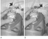 Figure 36-15
Sidestream sampling port placement. A,
To minimize the effects of breathing circuit dead space, attachment of the sampling
port should be as close to the patient as possible (arrow).
B, Placement of the port as shown (arrow)
can cause artifactual lowering of the end-tidal measurement.
Figure 36-15
Sidestream sampling port placement. A,
To minimize the effects of breathing circuit dead space, attachment of the sampling
port should be as close to the patient as possible (arrow).
B, Placement of the port as shown (arrow)
can cause artifactual lowering of the end-tidal measurement.
and are uncomfortable. Taping a piece of intravenous tubing close to the nostril
can provide an estimate of arterial PCO2
that is adequate for clinical purposes. Alternatively, an intravenous catheter can
be threaded into the common lumen of a pair of nasal O2
cannulas such
that the tip lies midway between the two nasal prongs. The extension tube normally
connected to the O2
source is tied off, and the intravenous catheter is
then connected to a sidestream capnometer.[170]
A commercially available version allows O2
administration while end-tidal
CO2
is continuously monitored (Divided Canula, Salter Labs, Arvin, CA).
Another device (Oridion, Needham, MA) samples exhalation from the mouth and nose
( Fig. 36-16
). Sampling
CO2
from a facemask, although adequate for monitoring respiratory rate,
produces measured PETCO2
values that are
significantly lower than PaCO2
.