|
|
|
|
|
|
|
|
|
|
|
|
|
|
|
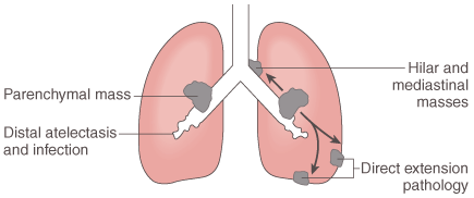
Figure 49-1
In patients with lung carcinoma, the chest radiographic
findings result from the presence of the tumor within the lung (parenchymal mass),
changes in the pulmonary parenchyma distal to a bronchus obstructed by the tumor
(atelectasis and infection), and spread of the tumor to extrapulmonary intrathoracic
sites (hilar and mediastinal masses and other direct extension pathology). (From
Benumof JL: Anesthesia for Thoracic Surgery. Philadelphia, WB Saunders, 1987.)

|
|
|
|
|
|
|
|
|
|
|
|
|
|