|
|
|
|
|
|
|
|
|
|
|
|
|
|
|
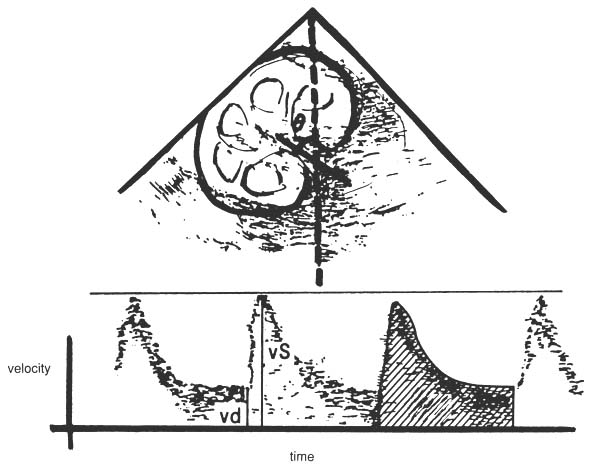
Figure 37-11
Renal duplex scan shows a sample volume superimposed
over a B-scan image for a typical normal velocity flow pattern from the main renal
artery at the hilum. theta, angle of incidence between transmitted ultrasound
signal and received ultrasound signal; vd, diastolic velocity; vS, systolic velocity.
|
|
|
|
|
|
|
|
|
|
|
|
|
|