 |
 |
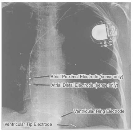
Figure 35-1
Pacemaker with one quadripolar lead that provides atrial
and ventricular sensing and ventricular pacing. This chest x-ray film shows a number
of features of a modern pacing system. The generator is located in the left pectoral
region. The single lead enters the subclavian vein under the clavicle but superficial
to the first rib (a common site for lead problems, although no problem is demonstrated
here). In this device, there are two electrodes in the right atrium that can provide
sensing to detect intrinsic atrial activity. The ventricular portion of the lead
shows the classic bipolar pattern, with a ring electrode just proximal to the tip
electrode, and these electrodes can be used for sensing intrinsic ventricular activity
and for depolarizing the ventricle. This is a ventricular pacing system with pacing
in the triggered and inhibited mode (VDD), and this configuration is placed into
patients with a functioning sinus node but a nonfunctioning atrioventricular node.
This system cannot be used to depolarize the atrium. Because the surface electrocardiogram
often demonstrates ventricular pacing that tracks the atrial activity, inspection
of the surface electrocardiogram typically produces an erroneous diagnosis of a dual
chamber (DDD) pacemaker.

 |
