 |
 |
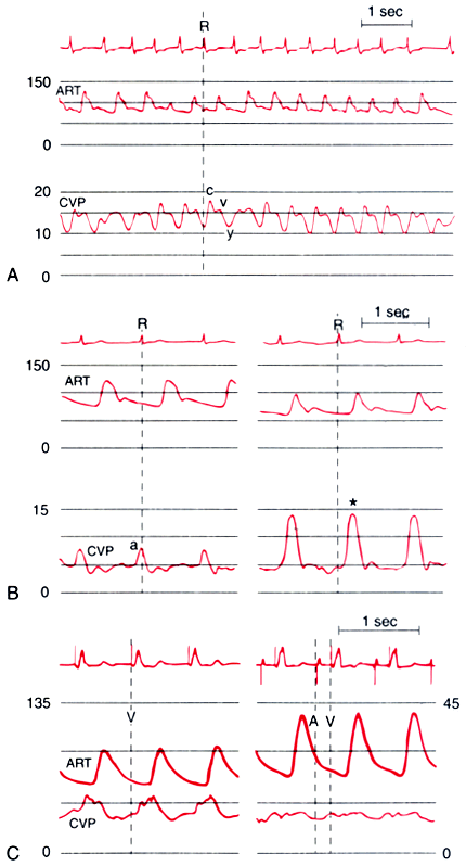
Figure 32-25
Central venous pressure (CVP) changes caused by cardiac
arrhythmias. A, Atrial fibrillation. Note the absence
of the a wave, a prominent c wave, and a preserved v wave and y descent. This arrhythmia
also causes variation in the electrocardiographic (ECG) R-R interval and left ventricular
stroke volume, which can be seen in the ECG and arterial pressure (ART) traces.
B, Isorhythmic atrioventricular dissociation. In
contrast to the normal end-diastolic a wave in the CVP trace (left
panel), an early systolic cannon wave is inscribed (asterisk,
right panel). The reduced ventricular filling accompanying this arrhythmia
causes decreased arterial blood pressure. C, Ventricular
pacing. Systolic cannon waves are evident in the CVP trace during ventricular pacing
(left panel). Atrioventricular sequential pacing
restores the normal venous waveform and increases arterial blood pressure (right
panel). The ART scale is shown on the left,
the CVP scale on the right. (Redrawn from
Mark JB: Atlas of Cardiovascular Monitoring. New York, Churchill Livingstone, 1998,
Figs. 14-1, 14-5, and 14-16.)

 |
