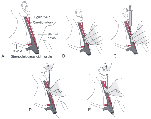
Figure 32-21
Technique for central venous cannulation of the right
internal jugular vein. A, Important surface landmarks
are identified. B, The course of the internal carotid
artery is palpated. C, The internal jugular vein
is punctured at the apex of the triangle formed by the two heads of the sternocleidomastoid
muscle, with the needle tip directed toward the ipsilateral nipple. D,
A guidewire is introduced through the thin-walled needle into the vein. E,
The central venous cannula is inserted over the guidewire while making sure that
the proximal end of the guidewire protrudes beyond the catheter and is controlled
by the operator.

