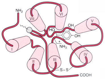
Figure 3-31
Schematic drawing of the three-dimensional structure
of the β2
-adrenergic receptor (a prototypical G protein-coupled receptor),
looking from the outside of the cell inward. Transmembrane domains (denoted by cylinders
and numbered with roman numerals) coalesce to form a binding pocket. The correct
orientation of the agonist norepinephrine is shown. Notice that agonist affinity
is determined by specific amino acids located in transmembranes III, IV, and VII.
These critical sites in transmembrane domains have been determined experimentally
using chimeric receptor and mutagenesis approaches combined with sophisticated computer-modeling
techniques. Ultimate confirmation of these structures will require crystallographic
data, an approach that is being attempted by several laboratories. (Adapted
from Stapelfeldt WH: The autonomic nervous system. In
Schwinn DA [ed]: Scientific Principles of Anesthesia, vol 2. Philadelphia, Current
Medicine, 1998.)