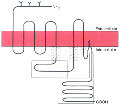
Figure 3-30
Schematic drawing of a G protein-coupled receptor. A
two-dimensional version of receptor structure is shown with seven transmembrane domains,
an extracellular amino (NH2
) terminus (with associated glycosylation sites
[Y]), an intracellular carboxyl (COOH) terminus, palmitoylated cysteine residue (crooked
line extending into the membrane), three extracellular loops, and four
intracellular loops. Major sites of G protein interactions with the receptor are
speckled; potential sites of phosphorylation in the third intracellular loop and
carboxyl terminus are enclosed in boxes.