|
|
|
|
|
|
|
|
|
|
|
|
|
|
|
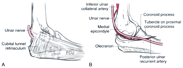
Figure 28-1
A, The retinaculum of
the cubital tunnel is depicted distinct from the aponeurosis of the flexor carpi
ulnaris (FCU) with which its distal margin blends. B,
The ulnar nerve and its primary blood supply in the proximal forearm, the posterior
ulnar recurrent artery, are superficial as they pass posteromedially to the tubercle
of the coronoid process. (Adapted from Warner MA: Perioperative neuropathies.
Mayo Clinic Proc 73:569, 571, 1998.)

|
|
|
|
|
|
|
|
|
|
|
|
|
|