|
|
|
|
|
|
|
|
|
|
|
|
|
|
|
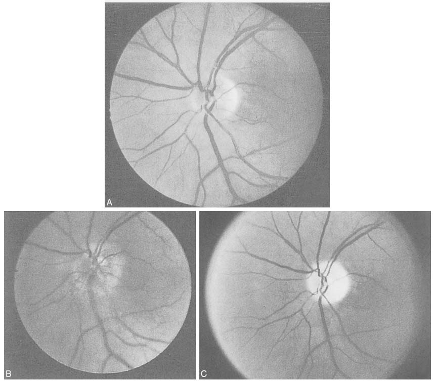
Figure 82-8
Funduscopic images showing the time course of changes
in anterior ischemic optic neuropathy (AION). A,
Normal fundus. B, Initial optic disk edema in AION.
C, Optic atrophy in the late stages. (From
Hayreh SS: Ischemic optic neuropathy. University of Iowa, Department of Ophthalmology
Internet Site. http://webeye.ophth.uiowa.edu/dept/aion/ion_fgD.jpg.)
|
|
|
|
|
|
|
|
|
|
|
|
|
|