|
|
|
|
|
|
|
|
|
|
|
|
|
|
|
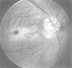
Figure 82-7
Funduscopic appearance of retinal vascular occlusion.
Note the pallor of the retina and the cherry-red spot, the clinical hallmark of
retinal arterial occlusion, visible in the fovea near the center of the picture.
The ischemic retina loses its normal transparency, and because the fovea is thinner
than the surrounding retina, the underlying choroid is visible as a cherry-red spot.
(From Ryan SJ: Retina, ed 2. St Louis, CV Mosby, 1995.)
|
|
|
|
|
|
|
|
|
|
|
|
|
|