|
|
|
|
|
|
|
|
|
|
|
|
|
|
|
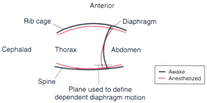
Figure 6-17
Diagram of a midsagittal section of the thorax while
awake (solid lines) and while anesthetized (red
lines) with a 1.2 minimum alveolar concentration (MAC) of halothane.
Chest wall configuration was determined using images of the thorax obtained by three-dimensional
fast computed tomography. (Adapted from Warner DO, Warner MA, Ritman EL:
Atelectasis and chest wall shape during halothane anesthesia. Anesthesiology 85:49,
1996.)

|
|
|
|
|
|
|
|
|
|
|
|
|
|