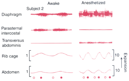
Figure 6-16
Representative record from one subject while awake and
during halothane anesthesia. The three upper tracings
are electromyograms. Lower tracings represent ribcage
and abdominal dimensions measured by respiratory impedance plethysmography. Open
and solid circles denote the beginning and end of inspiration, respectively.
Notice that the amplitudes of ribcage and abdominal excursions diminish during halothane
but that the relationship between their amplitudes is preserved. (From Warner
DO, Warner MA, Ritman EL: Human chest wall function while awake and during halothane
anesthesia. I. Quiet breathing. Anesthesiology 82:6, 1995.)