|
|
|
|
|
|
|
|
|
|
|
|
|
|
|
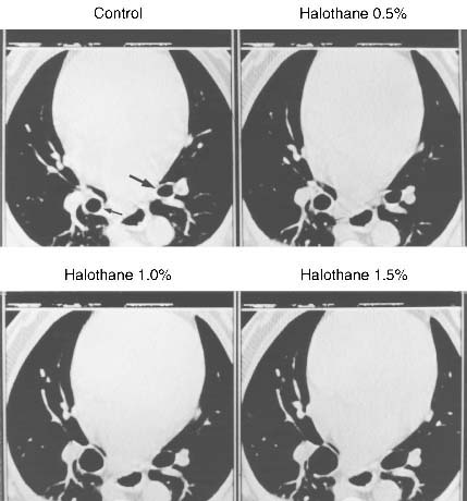
Figure 6-1
High-resolution computed tomography scans from one dog:
control (upper left), during 0.5% halothane (upper
right), during 1.0% halothane (lower left),
and during 1.5% halothane (lower right). Notice
the progressive dilation of the airways as indicated by the arrow.
(Adapted from Brown RH, Mitzner W, Zerhouni E, et al: Direct in vivo visualization
of bronchodilation induced by inhalational anesthesia using high-resolution computed
tomography. Anesthesiology 78:295, 1993.)
|
|
|
|
|
|
|
|
|
|
|
|
|
|