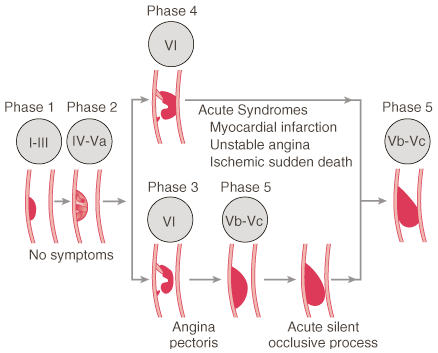
Figure 50-3
Description of lesion morphology and phases of progression
of coronary atherosclerosis in relation to clinical findings. Lipid accumulation,
phases I, II and III; thrombosis and hemorrhage, phases III and IV; and calcification
and fibrous tissue, phases IV and V. Roman numerals indicate lesion types: type
I to III lesions (early lesion) with isolated macrophage/foam cells (I), multiple
foam cell layers (II), or isolated extracellular lipids (III); type IV to Va lesions
(advanced lesions, atheromatous or fibrolipid plaques) with confluent extracellular
lipid pools (atheroma, IV) or fibromuscular tissue layers and atheroma (Va); type
VI lesions (advanced lesions, complicated plaques) with surface defects and/or hemorrhage
and/or thrombi deposition; type Vb to Vc lesions (advanced lesions) with calcifications
(Vb) or fibrous tissue (Vc). (From Fuster V, Gotto AM. Risk reduction.
Circulation 102:IV94–IV102, 2000.)