 |
 |
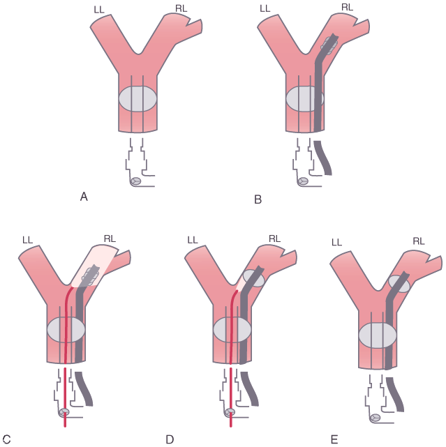
Figure 49-25
Lung separation with a single-lumen tube, fiberoptic
bronchoscope, and right lung bronchial blocker. The sequence of events is as follows.
A, A single-lumen tube is inserted and the patient
is ventilated. B, A bronchial blocker is passed alongside
the indwelling endotracheal tube. C, A fiberoptic
bronchoscope is passed through a self-sealing diaphragm in the elbow connecter to
the endotracheal tube and is used to place the bronchial blocker into the right main
stem bronchus under direct vision. D, The balloon
on the bronchial blocker is also inflated under direct vision and is positioned just
below the tracheal carina. E, The fiberoptic bronchoscope
is then removed. During the lower-panel sequence
(insertion and use of the fiberoptic bronchoscope ([C
to E]), the self-sealing diaphragm allows the patient
to continue to be ventilated with positive-pressure ventilation (around the fiberoptic
bronchoscope, but within the lumens of the endotracheal tube). LL, left lung; RL,
right lung. (From Benumof JL: Anesthesia for Thoracic Surgery. Philadelphia,
WB Saunders, 1987.)

 |
