 |
|
|
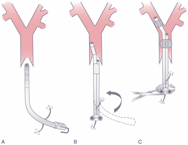
Figure 49-18
Schematic diagram depicting passage of the left-sided
double-lumen endotracheal tube in a supine patient. A,
The tube is held with the distal curvature concave anteriorly and the proximal curve
concave to the right and in a plane parallel to the floor. The tube is then inserted
through the vocal cords until the bronchial cuff passes the vocal cords. The stylet
is then removed. B, The tube is rotated 90 degrees
counterclockwise so that the distal curvature is concave anteriorly and the proximal
curvature is concave to the left and in a plane parallel to the floor. C,
The tube is inserted until either mild resistance to further passage is encountered
or the end of the common molding of the two lumens is at the teeth. Both cuffs are
then inflated, and both lungs are ventilated. Finally, one side is clamped while
the other side is ventilated and vice versa. (See the text for further explanation.)
(From Benumof JL: Anesthesia for Thoracic Surgery. Philadelphia, WB Saunders,
1987.)
|
|