Color Doppler Echocardiography
Point-by-point determination of blood flow velocities by PW Doppler
is too time consuming and does not reveal the instantaneous distribution of flow
velocities within the cross-sectional image that is required for many diagnostic
decisions. Color Doppler imaging was developed for this purpose. In color Doppler,
which is a form of PW Doppler, a color code is used to depict flow toward (red) and
away (blue) from the transducer; lighter and darker shades of red and blue respectively
denote relatively faster and slower velocities. Continuous color maps of flow are
superimposed on gray-scale cross-sectional echocardiograms. However, color Doppler
is generally a semiquantitative technique and, like PW Doppler, will alias (color
reversal) when the Nyquist limit is exceeded. Two aliasing patterns are easily recognized.
The first is "normal" aliasing in which the area of apparent flow reversal forms
one or more broad, relatively homogeneous color surfaces ( Plate
33-1
). Blood flow velocities within a normal heart often produce this
type of aliasing because they exceed the Nyquist limit for color Doppler (0.6 to
0.8 m/sec). The second type of aliasing results from disturbed or turbulent flow
within the heart (e.g., mitral regurgitation) and is never normal ( Plate
33-2
). When the ultrasonograph detects two different velocities
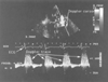 Figure 33-5
Continuous-wave Doppler measures high-velocity flow without
aliasing. Continuous-wave Doppler measurement of blood flow velocities in a mitral
valve orifice during four cardiac cycles is shown. At the top of the figure is a
still-frame image of the two-dimensional cross section used to position the Doppler
sample cursor (the diagonal white line). On the
bottom two thirds of the figure is the display in white of all the instantaneous
blood flow velocities (vertical axis) versus time (horizontal axis) occurring anywhere
along that cursor. The electrocardiogram provides timing, and the bold
horizontal line is the baseline (zero flow) for the flow velocities.
Flow velocities above this line are positive (i.e., toward the transducer) to a maximum
of 753 cm/sec. Flow velocities below this line are negative (i.e., away from the
transducer) to a maximum of -316 cm/sec. This tracing documents significant mitral
regurgitation (the positive systolic velocities) with a peak blood flow velocity
of approximately 5 m/sec (each white dot on the vertical
axis equals 100 cm/sec or 1 m/sec). LA, left atrium; LV, left ventricle. (From
Cahalan MK: Intraoperative Transesophageal Echocardiography. An Interactive Text
and Atlas. New York, Churchill Livingstone, 1997.)
Figure 33-5
Continuous-wave Doppler measures high-velocity flow without
aliasing. Continuous-wave Doppler measurement of blood flow velocities in a mitral
valve orifice during four cardiac cycles is shown. At the top of the figure is a
still-frame image of the two-dimensional cross section used to position the Doppler
sample cursor (the diagonal white line). On the
bottom two thirds of the figure is the display in white of all the instantaneous
blood flow velocities (vertical axis) versus time (horizontal axis) occurring anywhere
along that cursor. The electrocardiogram provides timing, and the bold
horizontal line is the baseline (zero flow) for the flow velocities.
Flow velocities above this line are positive (i.e., toward the transducer) to a maximum
of 753 cm/sec. Flow velocities below this line are negative (i.e., away from the
transducer) to a maximum of -316 cm/sec. This tracing documents significant mitral
regurgitation (the positive systolic velocities) with a peak blood flow velocity
of approximately 5 m/sec (each white dot on the vertical
axis equals 100 cm/sec or 1 m/sec). LA, left atrium; LV, left ventricle. (From
Cahalan MK: Intraoperative Transesophageal Echocardiography. An Interactive Text
and Atlas. New York, Churchill Livingstone, 1997.)
within the same small sample volume (because of disturbed flow), it displays a mixture
or mosaic of colors. These mosaics form jetlike configurations and are called "color
jets." Because color Doppler presents the spatial relationships between structure
and blood flow, it enhances the recognition of valvular abnormalities and intracardiac
shunts.
 |