Viscoelastic Measures of Coagulation
Initially developed in the 1940s, viscoelastic measures of coagulation
have undergone a resurgence in popularity. The unique aspect of viscoelastic monitors
lies in their ability to measure the entire spectrum of clot formation from early
fibrin strand generation through clot retraction and eventual fibrinolysis. The
thromboelastograph (TEG) (Haemoscope, Niles, IL), developed by Hartert in 1948,[840]
uses a small 0.35-mL blood sample placed into a disposable cuvette within the instrument.
The cuvette is maintained at a temperature of 37°C and continuously rotates
around an axis of approximately 5 degrees. A metal piston attached by a torsion
wire to an electronic recorder is lowered into the blood within the cuvette. As
clot formation occurs, the piston becomes enmeshed within the clot, and rotation
of the cuvette is transferred to the piston and electronic recorder.
Although variables derived from the TEG tracing do not coincide
directly with laboratory-based tests of coagulation, the TEG is capable of detecting
characteristic abnormalities in clot formation and fibrinolysis.[841]
Various parameters that describe the characteristics of clot formation and lysis
are inscribed by the TEG recorder. The R value (reaction time) measures the time
to initial clot formation (normal, 7.5 to 15 minutes). It is considered to be comparable
to the whole blood clotting time and may be accelerated by adding celite to the TEG
sample cuvette. The R value is prolonged by a deficiency of one or more plasma coagulation
factors. Maximum amplitude (MA) provides a measure of clot strength and may be decreased
by either qualitative or quantitative platelet dysfunction or decreased fibrinogen
concentration. Normal MA is 50 to 60 mm. The alpha angle and K (BiKoatugulierung
or coagulation) values measure the rate of clot formation and may be prolonged by
any factor slowing clot generation, such as plasma coagulation factor deficiency
or heparin anticoagulation ( Fig. 32-53
and Fig. 32-54
).
The Sonoclot (Sienco, Inc., Wheat Ridge, CO) provides an alternative
viscoelastic measure of coagulation. When compared with TEG, the Sonoclot immerses
a rapidly vibrating probe into a 0.4-mL sample of blood. As clot formation occurs,
impedance to probe movement through the blood increases and generates an altered
electrical signal and characteristic clot "signature." The Sonoclot may be used
to derive the ACT, as well as provide information regarding clot strength and clot
lysis.[842]
Both the TEG and Sonoclot generate characteristic diagrams by
translating the mechanical resistance encountered by the sensor as it moves through
the clotting blood sample. Measurements derived from these diagrams
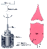 Figure 32-53
The functional components of the thromboelastograph consist
of a rotating cuvette, a suspended piston attached to a torsion wire, and a recorder.
As the blood sample clots, rotation of the cuvette is transferred to the torsion
wire and translated by the recorder as a characteristic tracing. Quantitative parameters
derived from the thromboelastograph trace are the reaction time (R), the BiKoatugulierung
value or coagulation time (K), the alpha angle (α), and maximum amplitude (MA).
See the text for greater detail. (Redrawn from Tuman K, Speiss B, McCarthy
R, et al: Effects of progressive blood loss on coagulation as measured by thromboelastography.
Anesth Analg 66:856, 1987.)
Figure 32-53
The functional components of the thromboelastograph consist
of a rotating cuvette, a suspended piston attached to a torsion wire, and a recorder.
As the blood sample clots, rotation of the cuvette is transferred to the torsion
wire and translated by the recorder as a characteristic tracing. Quantitative parameters
derived from the thromboelastograph trace are the reaction time (R), the BiKoatugulierung
value or coagulation time (K), the alpha angle (α), and maximum amplitude (MA).
See the text for greater detail. (Redrawn from Tuman K, Speiss B, McCarthy
R, et al: Effects of progressive blood loss on coagulation as measured by thromboelastography.
Anesth Analg 66:856, 1987.)
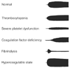 Figure 32-54
Characteristic thromboelastograph tracings.
Figure 32-54
Characteristic thromboelastograph tracings.
have been related to more traditional measures of coagulation such as the ACT.[843]
In addition, abnormal patterns have been associated with deficiencies in coagulation
factors and functional platelet abnormalities.[844]
[845]
One of the more common applications of the
TEG analyzer is the real-time detection of excessive fibrinolysis during liver transplantation.
The TEG may also be used to differentiate surgical bleeding from coagulopathy after
cardiac surgery.[846]
[847]
More widespread application of viscoelastic coagulation monitoring has been hindered
by the lack of specificity associated with abnormal findings and the qualitative
nature of assay interpretation. Recent computerization and automation of these instruments
have improved reproducibility of the measurements and made the results more quantitative.
Further modifications of viscoelastic measurement techniques, such as the addition
of heparinase to TEG samples to allow monitoring during cardiopulmonary bypass, may
lead to novel applications in the perioperative setting.[848]