Physiologic Considerations for Central Venous Pressure
Monitoring: Diastolic Pressure-Volume Relationships and Transmural Pressure
Cardiac filling pressures are monitored to estimate cardiac filling
volumes, which in turn determine the stroke output of the left and right ventricles.
According to the Frank-Starling principle, the force of cardiac contraction is directly
proportional to end-diastolic muscle fiber length at any given level of intrinsic
contractility or inotropy. This muscle fiber length or preload is proportional to
end-diastolic chamber volume. Even though it would be ideal to monitor cardiac chamber
volumes continuously in critically ill patients, this goal remains elusive in clinical
practice.
When a cardiac filling pressure is measured as a surrogate for
estimating cardiac volume, one must not assume that these two variables always change
in direct proportion or even in the same direction. In fact, the diastolic pressure-volume
relationship in cardiac muscle is not linear, but rather curvilinear, with a progressively
steeper slope at higher volumes ( Fig.
32-23
).[292]
[293]
This diastolic
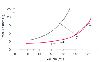 Figure 32-23
Ventricular diastolic pressure-volume relationship.
Along the flat portion of the curve, a 20-mL increase in ventricular volume causes
a small increase in ventricular pressure (A to B). In contrast, the same increase
in volume along the steep portion of the ventricular filling curve causes a marked
increase in filling pressure (C to D). Another problem associated with the use of
filling pressure as a surrogate for filling volume arises when shifts in the pressure-volume
relationship occur. At point C, ventricular volume is 100 mL and ventricular pressure
is 8 mm Hg. An increase in filling pressure to 15 mm Hg may accompany either increased
volume (D) or decreased volume (E). The latter occurs when ventricular compliance
changes and shifts the ventricular diastolic pressure-volume relationship up and
to the left. (Redrawn from Mark JB: Atlas of Cardiovascular Monitoring.
New York, Churchill Livingstone, 1998, Fig. 15-2.)
Figure 32-23
Ventricular diastolic pressure-volume relationship.
Along the flat portion of the curve, a 20-mL increase in ventricular volume causes
a small increase in ventricular pressure (A to B). In contrast, the same increase
in volume along the steep portion of the ventricular filling curve causes a marked
increase in filling pressure (C to D). Another problem associated with the use of
filling pressure as a surrogate for filling volume arises when shifts in the pressure-volume
relationship occur. At point C, ventricular volume is 100 mL and ventricular pressure
is 8 mm Hg. An increase in filling pressure to 15 mm Hg may accompany either increased
volume (D) or decreased volume (E). The latter occurs when ventricular compliance
changes and shifts the ventricular diastolic pressure-volume relationship up and
to the left. (Redrawn from Mark JB: Atlas of Cardiovascular Monitoring.
New York, Churchill Livingstone, 1998, Fig. 15-2.)
pressure-volume relationship is one limb of a pressure-volume loop that describes
the relationship between pressure and volume for the left or right ventricle during
an entire cardiac cycle. When a ventricle is operating along the flat portion of
its diastolic filling curve, a significant increase in filling volume or preload
results in a small increase in filling pressure. In contrast, the same increase
in filling volume causes a significant increase in filling pressure when the ventricle
is operating on the steep portion of its curve.[294]
An even more confusing situation arises when the diastolic pressure-volume relationship
of the ventricle changes, for example, with the onset of myocardial ischemia. Rather
than moving along the same diastolic pressure-volume curve, the ventricle now shifts
to a different, steeper curve where somewhat paradoxically, an increase in filling
pressure may accompany a decrease in filling volume.[295]
As a result, one cannot assume that a given measured change in cardiac filling pressure
reflects a proportional change in ventricular preload, and on occasion, diastolic
pressure and volume can change in opposite directions.[295]
[296]
The relationship between ventricular volume and filling pressure
depends on the portion of the pressure-volume curve over which the patient's heart
is operating and the shape or slope of the curve. Commonly termed ventricular compliance,
this change in pressure for a given change in volume (ΔP/ΔV) is actually
the reciprocal of compliance and is more accurately termed ventricular elastance,
distensibility, or stiffness.[297]
[298]
A patient with an abnormally stiff ventricle will have a greater change in end-diastolic
pressure for any given change in end-diastolic volume, and the converse is true for
a patient with an abnormally compliant ventricle. By definition, diastolic dysfunction
is present when ventricular pressure is abnormally elevated for any given ventricular
volume.
The ventricular diastolic pressure-volume relationship is influenced
by the intrinsic properties of the ventricle, such as the passive mechanical characteristics
of cardiac muscle, chamber geometry, and relaxation. In addition, external forces
exerted by the pericardium, the adjacent ventricle, the coronary vasculature, and
pleural pressure will further influence ventricular pressure-volume relationships.
[299]
[300]
[301]
[302]
One should not equate cardiac filling pressures
with filling volumes when patients are functioning over wide ranges of their diastolic
pressure-volume curve or under conditions in which diastolic stiffness is abnormal
or changing rapidly.
In general, all intravascular pressures measured in clinical practice
are referenced to ambient atmospheric pressure. (Indeed, the first step in pressure
transducer setup is to zero the transducer by exposing it to atmospheric pressure
and assigning this pressure a value of zero by pressing the zero pressure button
on the attached monitor. See "Technical Aspects of Direct Blood Pressure Monitoring.")
Thus, a cardiac filling pressure of 10 mm Hg is 10 mm Hg higher than ambient atmospheric
pressure. Does this pressure value accurately represent the distending force across
the cardiac chamber wall at end-diastole?
To answer this question, one needs to consider transmural pressure.
The cardiac chambers are all contained within the pericardium and thorax. Changes
in pressure in the structures surrounding the heart will influence
pressures recorded within the heart. Transmural pressure is the difference between
chamber pressure and juxtacardiac or pericardial pressure. This transmural pressure
determines ventricular preload, end-diastolic volume, or fiber length.[131]
[177]
The same measured filling pressure, referenced
to atmospheric pressure can be associated with markedly different transmural pressures
and chamber volumes, depending on whether juxtacardiac pressure is high or low.
Although juxtacardiac pressure can be ignored under some circumstances, marked alterations
in pleural and pericardial pressure occur commonly and must be considered when any
cardiac filling pressure is interpreted. Transmural pressure is always the pressure
of physiologic interest. Because juxtacardiac pressure is not measured routinely,
one always must consider that the measured central vascular pressure, referenced
to ambient atmospheric pressure, may be a poor estimate of transmural pressure.[294]
[303]
Cardiac filling pressures are measured directly from a number
of sites in the vascular system. CVP monitoring is the least invasive method, followed
by PAP monitoring and left atrial pressure monitoring. Proper interpretation of
all cardiac filling pressures requires knowledge of normal values for these pressures,
as well as pressures in the cardiac chambers and great vessels and other measured
and derived hemodynamic variables ( Table
32-7
).