|
|
|
|
|
|
|
|
|
|
|
|
|
|
|
Like the heart rate, blood pressure is a fundamental cardiovascular vital sign that describes the force driving tissue perfusion. Arterial blood pressure is the most important determinant of left ventricular afterload and, therefore, the workload of the heart. Consequently, frequent measurement of arterial blood pressure is a critical
Techniques for measuring blood pressure fall into two major categories: indirect Riva-Rocci cuff devices and direct arterial cannulation and pressure transduction. These methods differ most notably in terms of the physical signal being monitored and the level of invasiveness of their application. Although direct arterial blood pressure measurement is the reference standard against which other methods are compared, even this technique can yield spurious results. As a result, blood pressures measured in clinical practice with different techniques often yield significantly different values.[22] [23]
Most indirect methods of blood pressure measurement rely on a sphygmomanometer similar to the one first described by Riva-Rocci in 1896.[24] This apparatus included an arm-encircling inflatable elastic cuff, a rubber bulb to inflate the cuff, and a mercury manometer to measure cuff pressure.[25] Riva-Rocci described the measurement of systolic arterial blood pressure by determining the pressure at which the palpated radial arterial pulse disappeared as the cuff was inflated. The scientific rigor and attention to detail of Riva-Rocci's work are chronicled in a translation celebrating his original publications.[25]
A commonly used variation of the Riva-Rocci method is termed the return-to-flow technique. Whereas the Riva-Rocci technique recorded the pressure at which the pulse completely disappeared during cuff inflation, the return-to-flow method records the pressure at which the pulse reappears during cuff deflation. With this technique, systolic blood pressure can be estimated without a stethoscope by using only a cuff and manometer. When the patient has a finger pulse oximeter or indwelling arterial catheter in the ipsilateral arm, return to flow can be detected by reappearance of the plethysmographic or arterial pressure waveforms. Although return-to-flow methods provide a simple, rapid estimation of systolic blood pressure, they do not allow measurement of diastolic blood pressure.
To measure both systolic and diastolic arterial pressure, the most widely used intermittent manual method is the auscultatory technique, originally described by Korotkoff in 1905.[26] [27] Using a sphygmomanometer, cuff, and stethoscope, Korotkoff measured blood pressure by auscultation of the sounds generated by arterial blood flow. These sounds are a complex series of audible frequencies produced by turbulent flow, instability of the arterial wall, and shock wave formation as external occluding pressure on the artery is reduced.[28] The pressure at which the first Korotkoff sound is heard is generally accepted as systolic pressure (phase I). The sound character progressively changes (phases II and III), becomes muffled (phase IV), and is finally absent (phase V). Diastolic pressure is recorded at phase IV or V. However, phase V may never occur in certain pathophysiologic states such as aortic regurgitation.[29]
A fundamental shortcoming of the auscultatory method of blood pressure measurement is its reliance on blood flow to generate Korotkoff sounds. Pathologic or iatrogenic causes of decreased peripheral blood flow, such as cardiogenic shock or high-dose vasopressor infusion, can attenuate or obliterate sound generation and result in significant underestimation of blood pressure.[30] In contrast, low compliance of the tissues underlying the cuff, as encountered in a shivering patient, will require an excessively high cuff-occluding pressure and produce "pseudohypertension."[28] Patients with severe calcific arteriosclerosis have relatively noncompressible arteries, which is another circumstance wherein cuff blood pressures will overestimate the true intra-arterial blood pressure.[31]
Other common sources of error during intermittent manual blood pressure measurement include selection of an inappropriate cuff size and excessively rapid cuff deflation. The width of the blood pressure cuff should be 20% greater than arm diameter, and the cuff should be applied snugly after any residual air has been squeezed out. The pneumatic bladder inside the cuff should span at least half the circumference of the arm and be centered over the artery. Although too large a cuff will generally work well and produce little error, the use of cuffs that are too narrow will result in overestimation of blood pressure ( Fig. 32-2 ).[32] [33] The cuff deflation rate is another important variable that influences manual blood pressure measurement. The decrease in cuff pressure should proceed slowly enough for the Korotkoff sounds to be detected and properly assigned to the current pressure in the cuff. Failure to identify the initial Korotkoff sounds will result in a falsely low measurement of blood pressure. A deflation rate of 3 mm Hg/sec limits this source of error, and coupling the deflation rate to the heart rate—2 mm Hg/beat—has been found to improve accuracy further.[34]
Many limitations of manual intermittent blood pressure measurement have been overcome by automated noninvasive blood pressure (NIBP) devices, which are now used widely in medical care. By applying a single algorithm or
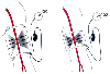 Figure 32-2
Effect of cuff size on manual blood pressure measurement.
An inappropriately small blood pressure cuff yields erroneously high values for
blood pressure because the pressure within the cuff is incompletely transmitted to
the underlying artery.
Figure 32-2
Effect of cuff size on manual blood pressure measurement.
An inappropriately small blood pressure cuff yields erroneously high values for
blood pressure because the pressure within the cuff is incompletely transmitted to
the underlying artery.
Most automated NIBP devices are based on oscillometry, a technique first described by von Recklinghausen in 1931.[35] In this method, variations in cuff pressure resulting from arterial pulsations during cuff deflation are sensed by the monitor and used to determine arterial blood pressure values. The pressure at which the peak amplitude of arterial pulsations occurs corresponds closely to directly measured mean arterial pressure (MAP),[36] [37] and values for systolic and diastolic pressure are derived from proprietary formulas that examine the rate of change of the pressure pulsations. Systolic pressure is identified as the pressure at which pulsations are increasing and are at 25% to 50% of maximum. Diastolic pressure is the most difficult value to determine by oscillometry and is commonly recorded when the pulse amplitude has declined from the peak value by 80% ( Fig. 32-3 ).[28]
Although oscillometry is used primarily in automated NIBP measurement, the same principles may be applied to determine blood pressure manually with a standard cuff and aneroid manometer. If the cuff is deflated slowly
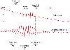 Figure 32-3
Comparison of blood pressure measurements by Korotkoff
sounds and oscillometry. Oscillometric systolic blood pressure is recorded at the
point where cuff pressure oscillations begin to increase, mean pressure corresponds
to the point of maximal oscillations, and diastolic pressure is measured when the
oscillations become attenuated. Note the correspondence between these measurements
and the Korotkoff sounds that determine auscultatory systolic and diastolic pressure.
(Redrawn from Geddes LA: Cardiovascular Devices and Their Applications.
New York John Wiley, 1984, Fig 34-2. Reprinted by permission of John Wiley &
Sons, Inc.)
Figure 32-3
Comparison of blood pressure measurements by Korotkoff
sounds and oscillometry. Oscillometric systolic blood pressure is recorded at the
point where cuff pressure oscillations begin to increase, mean pressure corresponds
to the point of maximal oscillations, and diastolic pressure is measured when the
oscillations become attenuated. Note the correspondence between these measurements
and the Korotkoff sounds that determine auscultatory systolic and diastolic pressure.
(Redrawn from Geddes LA: Cardiovascular Devices and Their Applications.
New York John Wiley, 1984, Fig 34-2. Reprinted by permission of John Wiley &
Sons, Inc.)
In clinical practice, oscillometric automated NIBP measurement has focused largely on pressures measured from the upper part of the arm. Although the cuff sizes recommended for NIBP measurement have followed the guidelines used for manual auscultatory techniques, when standard cuff sizes are used in critically ill patients, the pressure values recorded underestimate true intraarterial pressure, thus suggesting that smaller cuffs might be preferred for more accurate measurements in this setting.[38] On the other hand, if a patient's surgical procedure or medical condition requires that the cuff be applied to the calf, ankle, or thigh, an appropriately sized cuff must be used. Several investigators have proposed using neonatal-sized cuffs placed around the finger or thumb of an adult patient.[39] [40] [41] Although these alternatives may be applied in certain circumstances, their overall accuracy has not been widely validated and may not conform to accepted industry standards.[42]
Other techniques have been described for automated intermittent NIBP measurement. One method uses the Doppler principle to determine blood flow distal to an inflatable cuff,[43] a second involves photo-oscillometry to
Even though automated NIBP measurements have been shown to closely approximate directly measured arterial pressure in controlled clinical settings, [22] [33] [37] [46] numerous investigations underscore the disagreement that often exists when direct and indirect pressure measurements are compared, particularly when techniques are compared under changing hemodynamic conditions.[23] [47] [48] [49] Standards for performance of automated NIBP devices have been advanced by the American Association for the Advancement of Medical Instrumentation (AAMI) and the British Hypertension Society. AAMI standards require that a monitor record blood pressure to within 5 ± 8 mm Hg (mean ± standard deviation [SD]) prediction error with respect to the reference method.[44] However, the clinical performance of an NIBP monitor should be evaluated by other criteria, including the number of outlier values, duration of discrepancies, magnitude of individual errors, and performance under variable clinical conditions.[44]
Even though automated blood pressure measurement techniques are considered noninvasive and relatively safe, complications have been reported ( Table 32-1 ), including pain, petechiae and ecchymoses, limb edema, venous stasis and thrombophlebitis, peripheral neuropathy, and even compartment syndrome.[44] [50] [51] [52] These morbid events occur more often after prolonged periods of excessively frequent cuff inflation/deflation cycling and are due to trauma or impaired distal limb perfusion. Other factors that may contribute include cuff misplacement across a joint or repeated attempts to determine blood pressure in the presence of an artifact-producing condition such as involuntary muscle tremors.[53] Caution should be exercised when using these monitors in patients with depressed consciousness, preexisting peripheral neuropathies, arterial or venous insufficiency, or irregular cardiac rhythms[50] or in those receiving anticoagulant or thrombolytic therapy.[54]
Advances in microprocessor and servomechanical control technology
have enabled noninvasive techniques to
| Pain |
| Petechiae and ecchymoses |
| Limb edema |
| Venous stasis and thrombophlebitis |
| Peripheral neuropathy |
| Compartment syndrome |
Although several clinical investigators have demonstrated reasonable accuracy of finger blood pressure as a surrogate for intra-arterial pressure measurements, [61] a number of factors have precluded more widespread application of this technology. By definition, finger blood pressure monitoring records distal arterial pressure, which tends to be lower than brachial arterial pressure in elderly patients with atherosclerosis and higher than brachial pressure in young patients because of peripheral pulse wave amplification. Spasm of the finger arteries prevents accurate measurement in 5% of patients,[60] and some studies have noted substantial measurement errors in anesthetized and critically ill patients.[62] [63] [64] Unlike direct invasive arterial blood pressure monitoring, the finger pressure technique does not provide access for often-needed blood sampling. Because the "transducer" recording blood pressure with this method is the finger, the vertical height of the finger becomes an important determinant of the pressure recorded, just as transducer height is important with direct arterial pressure measurement. Finally, the potential for circulatory impairment of the distal end of the finger as a result of the constantly inflated cuff has been a cause for concern.[65]
Other automatic and continuous techniques have been used to measure blood pressure noninvasively. One such device reconstructs an arterial pressure waveform from measurements of arterial wall displacement after oscillometric calibration, but changes in arterial compliance appear to compromise the clinical performance of this instrument.[66] Another device uses pulse transit time as recorded from dual pulse oximeter probes placed on the ear and finger after oscillometric calibration from the contralateral arm, but it too has performed poorly in clinical settings, with indirectly monitored blood pressure changing in the opposite direction from direct intra-arterial pressure more than 30% of the time. [67] A third noninvasive method uses arterial tonometry, a variation of applanation tonometry in which a superficial artery (usually the radial) is compressed and partially flattened against the underlying bone.[68] [69] This flattened arterial surface
|
|
|
|
|
|
|
|
|
|
|
|
|