Positions for Orthopedic Surgery
Femoral neck fractures and midfemoral fractures needing open reduction
and internal fixation require different positions because of the need for surgical
access and for
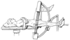 Figure 28-10
Femoral neck fractures can be managed in the supine position
on the fracture table. (Adapted from Martin JT: Lithotomy positions. In
Martin JT, Warner MA [eds]: Positioning in Anesthesia and Surgery, 3rd ed. Philadelphia,
WB Saunders, 1997, p 54.)
Figure 28-10
Femoral neck fractures can be managed in the supine position
on the fracture table. (Adapted from Martin JT: Lithotomy positions. In
Martin JT, Warner MA [eds]: Positioning in Anesthesia and Surgery, 3rd ed. Philadelphia,
WB Saunders, 1997, p 54.)
roentgenographic and fluoroscopic guidance ( Fig.
28-10
and Fig. 28-11
).
The lateral decubitus position typically is used for total-hip arthroplasty (see
Chapter 61
). The down hip
and leg are at risk during total-hip arthroplasty in the lateral decubitus position.
The orthopedic fracture table consists of a body section to support
the head and thorax, a sacral plate for the pelvis with a perineal post, and adjustable
footplates. The most important features of the table are the ability to maintain
traction on a lower extremity and to obtain surgical and fluoroscopic access. Because
the patients requiring this table are often in pain, anesthesia is usually induced
before the patient is moved to the table. If regional anesthesia is used, the fracture
side should be placed up to decrease the pain until the anesthetic takes affect.
After the patient has been transferred, the upper extremity on the fracture side
should be placed so that it does not interfere with surgical access to the fracture;
placing it across the chest directly or over an armboard is effective. Complications
from this position include brachial plexus injury, lower extremity compartment syndrome,
and pudendal nerve injury related to the perineal post.