Lateral Position
Thoracotomy and total-hip arthroplasty patients are usually placed
in the lateral position. The arm is placed perpendicular to the torso on a pillow
or on an overarm rest to support its weight ( Fig.
28-6
); the arm is often taped in this position. Care must be taken that
the tape does not impinge on the ulnar nerve at the elbow or on the radial
 Figure 28-6
The lateral position, showing the upper arm rest in position;
axillary roll, which supports the chest to free the axilla; and one type of leg positioning.
(Adapted from Day LJ: Unusual positions: Orthopedics: Surgical aspects.
In Martin JT [ed]: Positioning in Anesthesia and
Surgery, 2nd ed. Philadelphia, WB Saunders, 1987, p 226.)
Figure 28-6
The lateral position, showing the upper arm rest in position;
axillary roll, which supports the chest to free the axilla; and one type of leg positioning.
(Adapted from Day LJ: Unusual positions: Orthopedics: Surgical aspects.
In Martin JT [ed]: Positioning in Anesthesia and
Surgery, 2nd ed. Philadelphia, WB Saunders, 1987, p 226.)
nerve as it wraps around the radial groove in the upper third of the humerus. For
some thoracotomy procedures, a higher chest exposure is needed, and the arm is placed
above the shoulder plane. Special care must be taken to avoid plexus injury in these
situations ( Fig. 28-7
).
Tension on the brachial plexus can be reduced by bringing the arm into a more anterior
plane with the body. The lower area of the chest is generally supported with an
axillary roll, which often is a 1-L bag of intravenous fluid wrapped in a towel.
This places the weight of the chest on the rib cage and prevents the shoulder and
axilla from being compressed; compression of the axilla can lead to brachial plexus
injury in the down arm. Palpation of the arterial pulse in the down arm is sometimes
used as a measure of the adequacy of decompression. However, because the axillary
brachial plexus can be substantially compressed well before pulses are lost, this
method is probably insufficient evidence of safety. If the peripheral
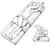 Figure 28-7
The lateral decubitus position for thoracotomy, showing
a more headward position of the arms to facilitate surgical exposure. (Adapted
from Lawson NW, Meyer DJ: Lateral positions. In
Martin JT, Warner MA [eds]: Positioning in Anesthesia and Surgery, 3rd ed. Philadelphia,
WB Saunders, 1997, p 134.)
Figure 28-7
The lateral decubitus position for thoracotomy, showing
a more headward position of the arms to facilitate surgical exposure. (Adapted
from Lawson NW, Meyer DJ: Lateral positions. In
Martin JT, Warner MA [eds]: Positioning in Anesthesia and Surgery, 3rd ed. Philadelphia,
WB Saunders, 1997, p 134.)
arm pulse is absent, substantial compression must have already occurred, and the
patient and chest roll should be repositioned.
Rhabdomyolysis much like that in a crush injury, arterial insufficiency
resulting in below-the-knee amputation, massive swelling of the thigh, and renal
failure associated with myoglobinuria have been reported.[29]
Use of the pulse oximeter to detect excessive pressure on the femoral triangle has
been suggested.[30]